Paperdoi:10.1097/01.hjh.0001197056.64038.c5
SPECKLE-BASED MEASUREMENT OF MICROVASCULAR FUNCTION
Journal of Hypertension 44(Suppl 1):e179, 2026
TL;DR
An imec conference abstract describes a finger speckle device using green and infrared lasers that probe different skin depths. Pressing on the finger made the shallow (green) speckle pulse shrink or vanish while the deeper infrared signal changed less, and a ratio of the two, a micro-to-macrovascular index, dropped by a median 55 %. The authors propose it as a step toward non-invasive measurement of microvascular function.
- Form factor: finger
Abstract
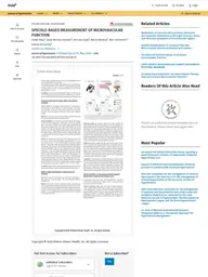
Objective: Microvascular function is compromised in many cardiovascular diseases, making it potentially suitable as an early marker for disease state. There are, however, no non-invasive and quantitative measurement methods of microvascular function available clinically. With our new dual-wavelength speckle sensing technology, we aim at enabling quantification of microvascular flow, comparing it to macrovascular function.Design and method: We developed a speckle device, which illuminates the finger with green and infrared laser light (Figure 1A). These wavelengths reach different depths of the skin, and therefore vasculature with different blood pressures (Figure 1B). A camera captures speckle images of the finger, from which speckle plethysmography (SPG) signals are calculated, which quantify the motion of scattering materials inside the finger. Measurements on 10 healthy volunteers (4 women, 27-60y) comprised placing the finger on the device during a resting phase and during applanation of the finger with a 400 g weight, resulting in a local change in transmural pressure. A micro-to-macrovascular index was calculated as the ratio of the green to infrared baseline/pulsatile components. Results: Representative examples of the SPG signals obtained (Figure 1C) show that the waveforms from the green and infrared light are comparable during rest. During applanation, the green SPG signal has a distinct change in pulse waveform, with a decrease or even complete disappearance of pulsation amplitudes (Subject 1 and 2, respectively). For the infrared signal however, the signal waveform changes less and even showed an increase in pulsation amplitude for Subject 1. The micro-to-macrovascular index decreases with 55 [3-81] % during applanation (median [range]), compared to a rest state. Conclusions: The strong decrease or complete disappearance of pulsations in the SPG signals from the green light during applanation suggests that the green signal measures vasculature under very low blood pressure. SPG signals from infrared light do not show this behaviour, indicating measurement of vasculature under higher pressure. Comparing these signals can describe the microvascular function compared to the macrovascular function. With this research, a step is made towards non-invasive and quantitative measurement of microvascular function.
Verbatim from OpenAlex (opens in a new tab) · Abstract via OpenAlex (publisher-deposited); © the authors/publisher
Results on ChatPPG leaderboards
All leaderboardsNo results from this paper on ChatPPG leaderboards yet. Browse the leaderboards.
Tasks & signals
Tasks
Signals
Keywords
How it works
- Wavelengths
- green laser, infrared laser
Cite this paper
Maas, E., Olazábal, J. H., van Kraaij, A., Barbeau, M., Verhoeven, M., & Hermeling, E. (2026). SPECKLE-BASED MEASUREMENT OF MICROVASCULAR FUNCTION. Journal of Hypertension 44:e179, 2026. https://doi.org/10.1097/01.hjh.0001197056.64038.c5
Maas, Esther, Jorge Herranz Olazábal, Alex van Kraaij, Marion Barbeau, Marc Verhoeven, and Evelien Hermeling. 2026. "SPECKLE-BASED MEASUREMENT OF MICROVASCULAR FUNCTION." Journal of Hypertension 44:e179, 2026. https://doi.org/10.1097/01.hjh.0001197056.64038.c5.
Maas, E., Olazábal, J. H., van Kraaij, A., Barbeau, M., Verhoeven, M. and Hermeling, E. (2026) 'SPECKLE-BASED MEASUREMENT OF MICROVASCULAR FUNCTION', Journal of Hypertension 44:e179, 2026. doi: 10.1097/01.hjh.0001197056.64038.c5.
Maas, Esther, et al. "SPECKLE-BASED MEASUREMENT OF MICROVASCULAR FUNCTION." Journal of Hypertension 44:e179, 2026, 2026. https://doi.org/10.1097/01.hjh.0001197056.64038.c5.
E. Maas, J. H. Olazábal, A. van Kraaij, M. Barbeau, M. Verhoeven, and E. Hermeling, "SPECKLE-BASED MEASUREMENT OF MICROVASCULAR FUNCTION," Journal of Hypertension 44:e179, 2026, 2026, doi: 10.1097/01.hjh.0001197056.64038.c5.
@article{maas2026specklebased,
  title         = {{SPECKLE-BASED MEASUREMENT OF MICROVASCULAR FUNCTION}},
  author        = {Maas, Esther and Olazábal, Jorge Herranz and van Kraaij, Alex and Barbeau, Marion and Verhoeven, Marc and Hermeling, Evelien},
  year          = {2026},
  journal       = {Journal of Hypertension 44:e179, 2026},
  doi           = {10.1097/01.hjh.0001197056.64038.c5}
}Added to the ChatPPG research hub