PPG Medical Meaning: What PPG Means in Clinical Practice
PPG in medical contexts means photoplethysmography — the optical technique used in pulse oximeters, hospital monitors, and wearables. Learn what it measures, what it shows clinicians, and how it is used.
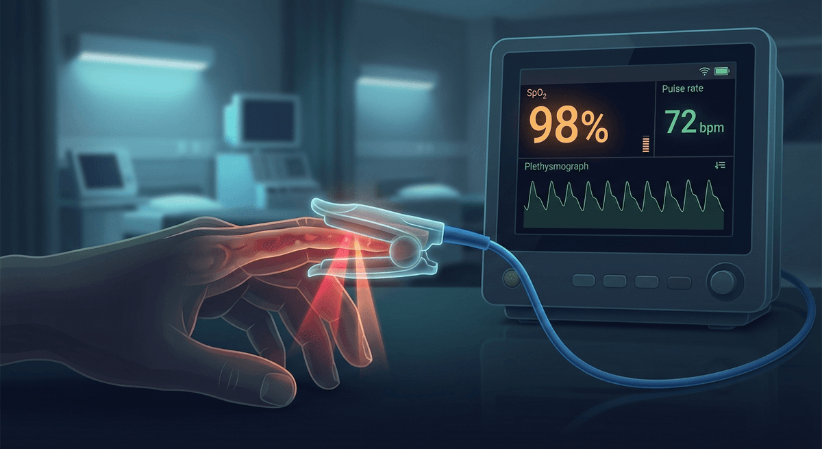
In medical contexts, PPG stands for photoplethysmography — the non-invasive optical technique that measures blood volume changes in tissue using light. PPG is central to clinical monitoring everywhere from emergency rooms and operating suites to post-surgical wards and outpatient cardiac telemetry programs. If you have ever had a fingertip probe clip attached during surgery or a hospital stay, you were being monitored with PPG.
The abbreviation appears in hospital monitor labels (the "Pleth" waveform is PPG), in nursing documentation (SpO₂ and pulse rate from the "PPG probe"), and increasingly in cardiology and sleep medicine as wearable PPG devices generate clinical data outside the hospital.
What PPG Measures in Clinical Settings
A medical PPG sensor shines light (typically red at 660 nm and infrared at 940 nm) through tissue and measures how much is absorbed. The pulsatile variation in absorption — rising with each heartbeat as blood volume increases, falling between beats — generates a continuous waveform called the photoplethysmogram.
From this waveform, clinical systems extract:
Pulse rate: The frequency of waveform peaks, expressed as beats per minute. Continuous pulse rate from PPG is the standard of care for perioperative and ICU monitoring.
Oxygen saturation (SpO₂): Using two wavelengths (red and NIR), the ratio of pulsatile signals encodes the proportion of hemoglobin carrying oxygen. Normal SpO₂ is 95–100%. Values below 90% are hypoxemia; below 85% represent severe hypoxemia requiring urgent intervention.
Perfusion index (PI): The ratio of pulsatile (AC) to non-pulsatile (DC) signal components, expressed as a percentage. A PI below 0.5–1% indicates poor peripheral perfusion — the PPG-derived numbers may be unreliable, and the patient may need circulatory assessment. PI above 1–5% is considered normal for most clinical contexts.
Pleth waveform morphology: The waveform shape itself carries clinical information about stroke volume, peripheral resistance, volume status, and cardiac rhythm. Experienced clinicians use waveform inspection alongside numeric readouts. See our detailed pleth waveform guide.
Respiratory rate (from PPG): PPG waveform amplitude is modulated by respiration — amplitude increases slightly with expiration and decreases with inspiration (due to intrathoracic pressure effects on venous return). Some advanced monitors compute respiratory rate from this amplitude modulation and from baseline wander in the PPG signal.
PPG in the Operating Room and ICU
Perioperative PPG monitoring has been standard since the 1980s, when the pulse oximeter was introduced as a patient safety technology. The story of the pulse oximeter's adoption is one of the most impactful device introductions in anesthesia history — before its availability, intraoperative hypoxemia was often detected late or not at all.
In modern operating rooms, the PPG sensor (via the SpO₂ probe) provides:
- Continuous SpO₂ with alarm limits (typically 92–94% lower alarm threshold)
- Continuous pulse rate as a backup to ECG-derived heart rate
- Pleth waveform display for hemodynamic assessment
- Perfusion index for peripheral perfusion monitoring
- Plethysmographic variability index (PVI): Dynamic measure of respiratory modulation of the PPG waveform, used to predict fluid responsiveness in mechanically ventilated patients. PVI >13–15% suggests the patient may respond to a fluid bolus.
In the ICU, PPG is used similarly, with the addition of longer-duration monitoring for:
- Detecting early hemodynamic deterioration through waveform amplitude changes
- Sepsis-related perfusion monitoring
- Sleep quality assessment in long-term ICU patients (some centers)
- Post-extubation SpO₂ monitoring
PPG Probe Types Used in Hospitals
Different clinical situations call for different probe placements:
Fingertip clip (most common): Spring-loaded clip over the fingertip pad. Transmission mode — LED and detector on opposite sides. Best signal quality in well-perfused patients. Fails with poor peripheral circulation, nail polish, dark nails, or cold fingers.
Forehead reflectance sensor: Adhesive sensor on forehead, reflectance mode. More reliable in hypotension, vasoconstriction, and cold environments. Used in ICU and operating room when fingertip is unreliable. Not affected by nail polish.
Earlobe clip: Clip sensor on earlobe. Good perfusion, minimal motion artifact. Alternative when finger probe fails. Less affected by peripheral vasoconstriction than fingertip.
Neonatal and pediatric sensors: Wrap-around adhesive sensors for fingers, toes, or feet. Sized for small patients; designed for thin tissue sites. Neonatal sensors require lower LED intensity settings to avoid skin damage.
Reusable vs. disposable: ICU often uses adhesive disposable sensors for hygiene; reusable clip probes are standard for shorter monitoring periods in surgical suites.
PPG in Clinical Cardiology
Beyond bedside monitoring, PPG has entered clinical cardiology workflows through consumer wearable data, dedicated cardiac monitoring devices, and clinical-grade ambulatory monitoring systems.
Ambulatory cardiac monitoring: Companies like iRhythm (Zio patch), BioTel Heart, and Preventice (BodyGuardian) deploy multi-day to multi-week monitoring patches that include PPG alongside ECG. PPG provides continuous hemodynamic context for arrhythmias detected on ECG.
AF detection via wearables: Since 2018, FDA-cleared wearable devices (Apple Watch, Withings ScanWatch) have been submitting PPG-based AF notifications to patients, who then present to cardiologists for confirmation. Multiple academic cardiology centers have reported that 20–40% of wearable-triggered cardiology consultations result in a new AF diagnosis. This has meaningfully changed early AF detection patterns.
Sleep cardiology: PPG-based SpO₂ monitoring during sleep is used for home sleep apnea testing (HSAT). Several FDA-cleared HSAT devices (WatchPAT, Nox T3, ResMed) use PPG-derived SpO₂ and peripheral arterial tone (PAT) as primary diagnostic signals. This has dramatically expanded access to sleep apnea diagnosis outside of formal sleep labs.
PPG in Emergency Medicine
Emergency physicians encounter PPG in several contexts:
Trauma resuscitation: PI and waveform amplitude during trauma resuscitation provide rapid assessment of peripheral perfusion and volume status. A rising PI after fluid administration indicates improving perfusion. Waveform variation on mechanical ventilation guides further fluid therapy.
Sepsis identification: Early septic vasodilation produces a characteristic high-amplitude, bouncy PPG waveform. As sepsis progresses to septic shock, waveform amplitude falls with dropping blood pressure. Bedside PPG provides a continuous hemodynamic indicator.
Overdose monitoring: SpO₂ monitoring is essential in opioid overdose and respiratory depression. PPG SpO₂ plus waveform monitoring allows detection of hypoxemia and hemodynamic compromise.
Carbon monoxide poisoning: Standard PPG SpO₂ cannot detect carboxyhemoglobin (COHb). Regular pulse oximeters read SpO₂ falsely normal in CO poisoning because COHb absorbs red light similarly to oxyhemoglobin. Multi-wavelength CO-oximeters (Masimo Rad-57) use additional wavelengths to detect COHb. Physicians must know this limitation — a normal SpO₂ in a suspected CO poisoning patient requires CO-oximetry or laboratory co-oximetry.
Understanding PPG in Your Hospital Monitor
If you or a family member is in the hospital and you are looking at the monitor, the "Pleth" or "SpO₂ wave" is the PPG waveform. Here is how to interpret it at a glance:
- Regular, medium-height peaks: Normal cardiac rhythm and adequate peripheral perfusion
- Irregular peaks of varying height: Consider arrhythmia (AF, frequent ectopics)
- Very small, flat waveform: Poor perfusion — either the probe is loose, or the patient has circulatory compromise
- Jagged, noisy waveform: Motion artifact or electrical interference — the SpO₂ number may be unreliable
- Regular peaks with rhythmic amplitude variation: Normal respiratory modulation (healthy finding in spontaneously breathing patients)
Frequently Asked Questions
What does PPG stand for in medical terms? PPG stands for photoplethysmography. The adjective form is "photoplethysmographic" (as in "photoplethysmographic sensor" or "PPG probe"). The measured signal is the photoplethysmogram.
Is a pulse oximeter the same as a PPG monitor? A pulse oximeter uses PPG as its underlying measurement technology. The SpO₂ value is derived by analyzing the PPG signal at two wavelengths. So a pulse oximeter is a specialized PPG monitor configured for oxygen saturation measurement. Standalone PPG monitors (sometimes called plethysmographs) provide the waveform without necessarily computing SpO₂.
What is a normal PPG reading in hospital? "PPG reading" usually refers to SpO₂ and pulse rate. Normal SpO₂: 95–100% in adults breathing room air. Normal pulse rate: 60–100 bpm at rest. Normal perfusion index: 0.5–15% (varies by body site; fingertip PI of 1–5% is typical in a well-perfused resting adult).
Why does my PPG probe keep alarming in the hospital? Common causes: low perfusion (check if fingers are cold — try warming them or moving the probe to the forehead or earlobe), motion artifact (movement disrupts the waveform), nail polish or artificial nails (dark colors interfere with light transmission), probe not secured properly. Persistent low SpO₂ alarms should always be clinically evaluated, not dismissed.
Can PPG be used to measure hemoglobin? Experimental multi-wavelength PPG systems can estimate total hemoglobin (SpHb) non-invasively. Masimo Rainbow SET uses 8 wavelengths to estimate SpHb, SpMet (methemoglobin), SpCO (carboxyhemoglobin), and others. Accuracy for SpHb is moderate (±1–2 g/dL versus laboratory co-oximetry) and is used as a trend indicator rather than a precise measurement. See our guide to PPG SpO₂ accuracy limitations.
Is PPG accurate in patients with dark skin tones? Standard dual-wavelength PPG (660/940 nm) for SpO₂ can overestimate SpO₂ in patients with darker skin, particularly at lower true saturations. This was extensively documented by Sjoding et al. (2020, NEJM) and has prompted FDA guidance requiring manufacturers to study oximeter accuracy across skin tone ranges. Newer multi-wavelength designs reduce but do not eliminate this bias.
References
-
Jubran, A. (2015). Pulse oximetry. Critical Care, 19, 272. https://doi.org/10.1186/s13054-015-0984-8
-
Severinghaus, J. W., & Honda, Y. (1987). History of blood gas analysis. VII. Pulse oximetry. Journal of Clinical Monitoring, 3(2), 135–138. https://doi.org/10.1007/BF00858177
-
Sjoding, M. W., Dickson, R. P., Iwashyna, T. J., Gay, S. E., & Valley, T. S. (2020). Racial bias in pulse oximetry measurement. New England Journal of Medicine, 383(25), 2477–2478. https://doi.org/10.1056/NEJMc2029240
-
Cannesson, M., Attof, Y., Rosamel, P., et al. (2008). Respiratory variations in pulse oximetry plethysmographic waveform amplitude to predict fluid responsiveness. Anesthesiology, 106(6), 1105–1111. https://doi.org/10.1097/ALN.0b013e318173f092