What Is Pulse Wave Velocity? Normal Ranges, Clinical Thresholds, and PPG Estimation
Plain-language explanation of pulse wave velocity, normal ranges by age, clinical risk thresholds, and how PPG wearables estimate PWV from pulse transit time.
Pulse wave velocity (PWV) is the speed at which the pressure wave generated by each heartbeat travels through your arteries. It is measured in meters per second (m/s) and serves as the reference standard for assessing arterial stiffness. In healthy young adults, PWV through the aorta typically falls between 6 and 8 m/s. As arteries stiffen with age, that speed climbs to 10 to 12 m/s or higher. A carotid-femoral PWV above 10 m/s is recognized by the European Society of Hypertension and the European Society of Cardiology as a marker of increased cardiovascular risk. PWV is simple in concept, clinically powerful, and increasingly measurable through wearable PPG devices that estimate it from pulse transit time.
Your heart does this roughly 100,000 times per day. Each contraction sends a wave of pressure into the aorta, and that wave ripples outward through every artery until it reaches the smallest vessels in your fingers and toes. The speed of that ripple is pulse wave velocity.
The Garden Hose Analogy
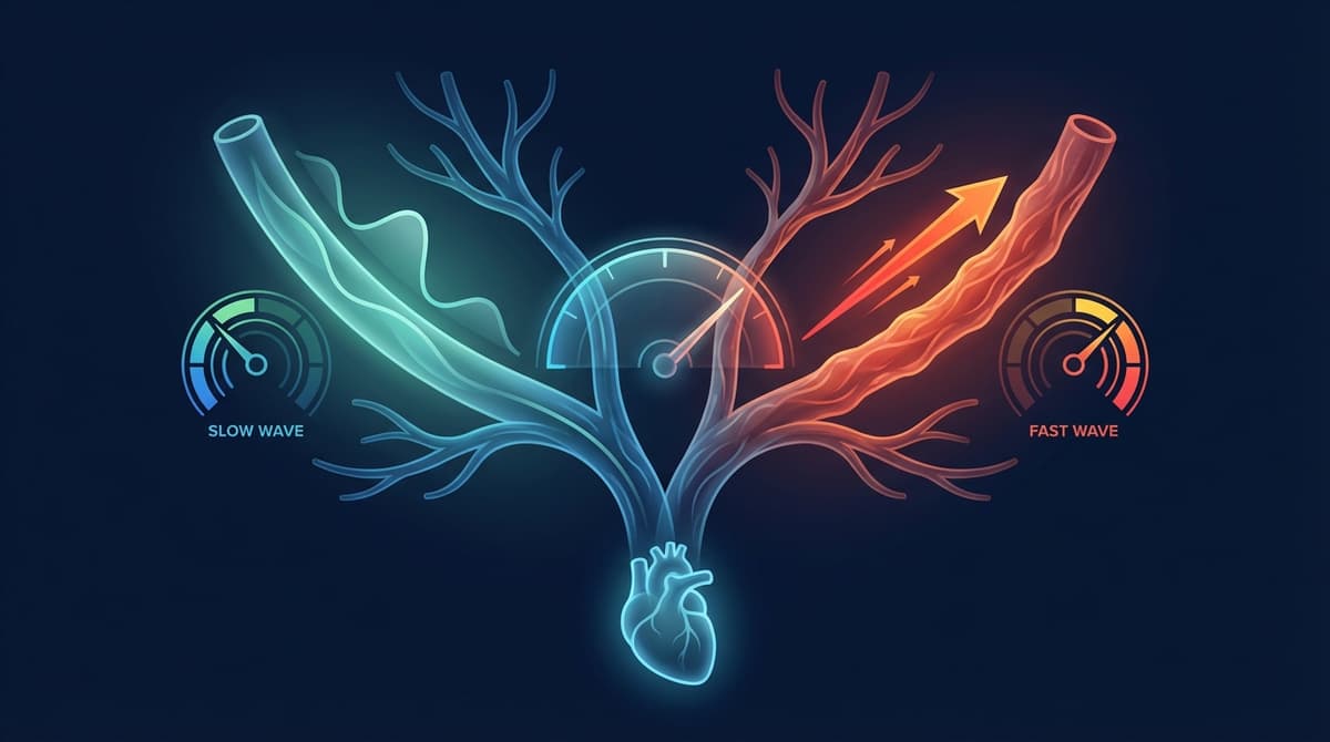
Imagine two garden hoses. One is soft, flexible rubber. The other is rigid PVC pipe. If you create a pressure pulse at one end of the soft hose, the bulge moves slowly because the hose wall stretches to absorb energy as the wave passes. In the stiff pipe, there is nowhere for that energy to go except forward, so the wave races through much faster.
Your arteries work the same way. Young, healthy arteries are elastic. They stretch when the heart pushes blood into them, store that energy briefly, then recoil to push blood forward during diastole (the resting phase between heartbeats). This cushioning effect is called the Windkessel function, and it smooths out the pulsatile flow from the heart into a steadier flow downstream.
When arteries stiffen, whether from aging, high blood pressure, diabetes, or other factors, they lose that elastic cushion. The pressure wave passes through faster. PWV goes up. That faster wave also bounces back from branch points and peripheral vessels sooner, arriving back at the heart while it is still pumping rather than while it is relaxed. The result is higher systolic blood pressure, increased cardiac workload, and reduced coronary perfusion. This is why arterial stiffness matters, and why measuring it through PWV has become a target of cardiovascular research and clinical practice.
How PWV Is Measured
The core measurement is straightforward. You need two things: a known distance between two arterial sites and the time it takes the pressure wave to travel between them. Divide distance by time and you have velocity.
PWV = distance / transit time
The gold standard method is carotid-femoral PWV (cfPWV). A pressure sensor is placed on the neck over the carotid artery and another on the groin over the femoral artery. Both sensors record the local pulse waveform, and the time delay between the two waveforms gives the transit time. The surface distance between the two sites, corrected for body geometry, provides the path length.
Other methods exist. Brachial-ankle PWV (baPWV), widely used in Japan and other Asian countries, places blood pressure cuffs on the upper arm and ankle. Finger-toe PWV uses optical sensors at the extremities. Each method measures a different arterial segment and produces different absolute values, which is why specifying the measurement method matters when comparing results. For a deeper comparison of these approaches, see our guide on pulse wave velocity measurement methods.
Normal Ranges by Age
PWV increases steadily with age, even in healthy individuals, because arteries gradually lose elastin and accumulate collagen. The Reference Values for Arterial Stiffness Collaboration, which pooled data from over 16,000 subjects across 13 centers, established the following approximate carotid-femoral PWV ranges for healthy individuals without cardiovascular risk factors (DOI: 10.1093/eurheartj/ehq165):
| Age Group | Mean cfPWV (m/s) | Typical Range (m/s) |
|---|---|---|
| Under 30 | 6.2 | 5.3 to 7.1 |
| 30 to 39 | 6.5 | 5.5 to 7.7 |
| 40 to 49 | 7.2 | 6.0 to 8.6 |
| 50 to 59 | 8.3 | 6.8 to 10.0 |
| 60 to 69 | 10.3 | 8.0 to 13.1 |
| 70 and older | 10.9 | 8.5 to 14.6 |
A few points stand out. The spread within each age group is wide. A 55-year-old with a cfPWV of 7.5 m/s has arteries behaving like those of someone decades younger, while a 55-year-old at 11 m/s is tracking above expectations. These values are for carotid-femoral PWV specifically; brachial-ankle values run 20 to 50% higher because the path includes stiffer muscular arteries. Also, the acceleration after age 50 is striking. PWV does not climb in a straight line; it curves upward as cumulative pressure damage compounds.
The 10 m/s Clinical Threshold
The 2018 ESC/ESH Guidelines for the Management of Arterial Hypertension identify a carotid-femoral PWV exceeding 10 m/s as evidence of hypertension-mediated organ damage (DOI: 10.1093/eurheartj/ehy339). This threshold is not arbitrary. It emerged from multiple large prospective studies showing that individuals crossing this line face significantly higher rates of cardiovascular events, independent of blood pressure itself.
What does "independent of blood pressure" mean in practice? It means that two people can have the same blood pressure reading, say 140/90 mmHg, but the one with a PWV of 12 m/s faces higher risk than the one at 8 m/s. Blood pressure measures force at a single moment. PWV reflects the accumulated structural state of the arterial wall. It captures damage that blood pressure alone cannot.
The 10 m/s cutoff applies to carotid-femoral measurements using the 80% distance correction recommended by the ARTERY Society. Using a different distance method or a different arterial segment will shift the threshold. This is a common source of confusion in clinical practice and research, and it underscores why standardization matters.
What Affects Pulse Wave Velocity
PWV is influenced by a mix of non-modifiable and modifiable factors.
Non-Modifiable Factors
Age is the strongest determinant. The progressive fragmentation of elastin fibers and accumulation of collagen in arterial walls is a universal feature of vascular aging. By age 70, the aorta may contain less than half the functional elastin it had at age 20.
Genetics play a role as well. Some individuals have inherently stiffer or more compliant arteries for their age, and family studies suggest that 30 to 50% of the variance in PWV is heritable.
Modifiable Factors
Hypertension is the most potent accelerator of arterial stiffening. Chronically elevated pressure damages elastic fibers and triggers smooth muscle remodeling. The relationship is bidirectional: stiff arteries raise blood pressure, and high blood pressure stiffens arteries further. This creates a vicious cycle that is difficult to break without intervention.
Diabetes increases PWV through multiple pathways. Hyperglycemia promotes the formation of advanced glycation end products (AGEs) that cross-link collagen fibers, making the arterial wall rigid. Insulin resistance also drives inflammation and endothelial dysfunction, both of which contribute to stiffening.
Smoking damages the endothelium and promotes oxidative stress, accelerating elastin degradation. Studies consistently show that smokers have higher PWV than age-matched nonsmokers. The good news: quitting smoking leads to measurable reductions in arterial stiffness within months.
Physical inactivity is associated with higher PWV, while regular aerobic exercise reduces it. A meta-analysis found that moderate-intensity aerobic training for 8 to 26 weeks reduced cfPWV by an average of 0.7 m/s. Epidemiological data suggest that each 1 m/s increase in cfPWV is associated with a 14 to 15% increase in cardiovascular events.
Diet also matters. High sodium intake increases arterial stiffness acutely and chronically. Diets rich in fruits, vegetables, and omega-3 fatty acids are associated with lower PWV. For more on vascular age tracking, see our article on PPG-based vascular age assessment.
How PPG Wearables Estimate PWV
Traditional PWV measurement requires dedicated equipment, a trained operator, and a clinical setting. PPG-based estimation aims to change that by using the optical sensors already built into smartwatches, fitness bands, and pulse oximeters.
Pulse Transit Time as a Proxy
The most common approach uses pulse transit time (PTT), the time interval between a cardiac event and the arrival of the pulse wave at a peripheral PPG sensor. In a typical setup, an ECG or chest-mounted sensor detects the electrical trigger of the heartbeat (the R-wave), while a wrist or finger PPG sensor detects the arriving pulse wave. The delay between these two events approximates the time the pressure wave took to travel from the heart to the sensor site.
From PTT, PWV can be estimated if the path length is known or approximated from body measurements:
PWV = estimated arterial path length / PTT
This is the principle behind many research prototypes and some commercial devices. For a detailed look at this approach, see our guide on pulse transit time and blood pressure estimation.
Single-Site PPG Estimation
An even more accessible approach extracts PWV-related information from a single PPG sensor, avoiding the need for a separate ECG reference. This works because the shape of the PPG waveform encodes information about arterial stiffness. Features like the stiffness index (body height divided by the time between the systolic and diastolic peaks), the augmentation index, and second-derivative features all correlate with PWV.
Machine learning models trained on paired PPG and reference PWV measurements can combine multiple waveform features to estimate PWV with moderate accuracy. Published studies report correlations of r = 0.6 to 0.8 between PPG-estimated and reference cfPWV, with mean absolute errors typically in the range of 1.0 to 2.0 m/s.
Limitations and Progress
PPG-based PWV estimation is promising but not yet clinically validated for standalone use. Several challenges remain:
- Path length uncertainty. Estimating arterial distance from body height or arm length introduces error that directly propagates into the PWV calculation.
- Pre-ejection period. PTT includes the time between the electrical trigger and actual ejection of blood (the pre-ejection period, or PEP), which is not part of arterial transit and varies with cardiac contractility.
- Motion artifacts. Wrist PPG signals are noisy during daily activities, and motion artifacts can distort the waveform features used for estimation.
- Calibration drift. Individual differences in anatomy, skin pigmentation, and sensor placement mean that models calibrated on one population may not generalize well.
Despite these challenges, the field is advancing rapidly. Multi-sensor fusion, improved signal processing algorithms, and larger training datasets are steadily improving accuracy. The long-term vision is continuous, passive PWV monitoring that flags arterial stiffening early enough for lifestyle intervention to make a difference. For a broader perspective on how PPG signals relate to aortic stiffness, see our article on PPG as an aortic stiffness biomarker.
Why PWV Matters for You
PWV is one of the few metrics that captures the structural health of your blood vessels, not just what your heart is doing right now but how well the plumbing has held up over decades. A high PWV for your age is a warning sign that arrives years before a heart attack or stroke. A low PWV is reassurance that your cardiovascular system is aging well.
As wearable PPG devices get better at estimating PWV, this metric will move from the vascular lab to the wrist, giving millions of people a window into their arterial health that was previously available only through specialized testing.
Frequently Asked Questions
What is a normal pulse wave velocity for my age?
For healthy adults, carotid-femoral PWV averages about 6 to 7 m/s in your 20s and 30s, 7 to 8 m/s in your 40s, 8 to 10 m/s in your 50s, and 10 to 11 m/s in your 60s and 70s. Individual values vary based on genetics, fitness level, and risk factors. A value below the average for your age group is generally favorable.
Is a pulse wave velocity above 10 m/s dangerous?
A cfPWV above 10 m/s is classified as a marker of increased cardiovascular risk by the ESH/ESC. It does not mean immediate danger, but it signals that your arteries are stiffer than ideal and your risk of heart attack, stroke, and other events is elevated. It is a reason to discuss managing risk factors like blood pressure, blood sugar, cholesterol, and physical activity with your doctor.
Can you lower pulse wave velocity?
Yes, PWV is modifiable. Regular aerobic exercise, blood pressure control through medication or lifestyle changes, smoking cessation, and dietary improvements have all been shown to reduce PWV in clinical studies. The effect sizes are modest, typically 0.5 to 1.5 m/s, but even small reductions carry meaningful risk reduction. The key is sustained effort over months and years rather than short-term changes.
How do smartwatches measure pulse wave velocity?
Most smartwatches estimate PWV indirectly rather than measuring it directly. They use the PPG optical sensor to capture pulse waveforms and extract features related to arterial stiffness, such as the timing of reflected waves, pulse transit time (when combined with an ECG reference), and waveform shape features. Machine learning models then translate these features into a PWV estimate. The accuracy is improving but is not yet equivalent to clinical devices like SphygmoCor or Complior.
What is the difference between pulse wave velocity and blood pressure?
Blood pressure measures the force of blood against artery walls at a single moment (for example, 120/80 mmHg). PWV measures how fast the pressure wave travels through your arteries (in m/s). Blood pressure fluctuates throughout the day with posture, stress, and activity. PWV reflects the underlying structural stiffness of the arterial wall, which changes slowly over months and years. Two people with identical blood pressure can have very different PWV values, and the one with higher PWV faces greater long-term risk.
Is pulse wave velocity the same as heart rate?
No. Heart rate is how many times your heart beats per minute. Pulse wave velocity is how fast the pressure wave from each beat travels through your arteries. A person with a heart rate of 70 bpm could have a PWV of 7 m/s (healthy, compliant arteries) or 13 m/s (stiff arteries). While heart rate and PWV are both extracted from pulse signals, they measure entirely different things. Heart rate reflects cardiac rhythm; PWV reflects vascular health.
Does pulse wave velocity change throughout the day?
PWV fluctuates modestly with blood pressure, heart rate, and sympathetic activity. It tends to be slightly higher in the morning, during exercise, and during stress. However, the resting baseline reflects your underlying arterial stiffness and changes only gradually over weeks to months. Clinical assessments are performed under standardized resting conditions to minimize this variability.