Pulse Wave Velocity Measured by PPG: What It Is, Normal Values, and Clinical Meaning
Pulse wave velocity (PWV) is the speed at which a pressure wave travels through your arteries after each heartbeat. A higher PWV means stiffer arterie...
Pulse wave velocity (PWV) is the speed at which a pressure wave travels through your arteries after each heartbeat. A higher PWV means stiffer arteries — and stiffer arteries mean higher cardiovascular risk. PPG sensors in modern wearables can estimate PWV non-invasively, making this once-clinical measurement available to anyone with a smartwatch.
What Is Pulse Wave Velocity?
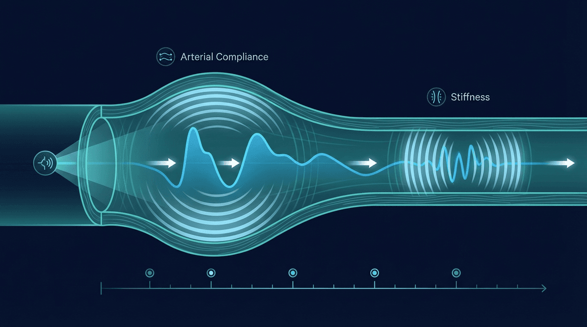
When your heart contracts and ejects blood, it creates a pressure pulse that propagates along the arterial wall — much like a wave along a stretched rope. The speed of that wave depends on the mechanical stiffness of the arterial wall: stiff walls transmit pressure faster, compliant walls slower.
PWV is expressed in meters per second (m/s) and calculated as:
PWV = Distance / Transit Time
Where distance is the arterial path length between two measurement sites, and transit time is the delay between the pulse arriving at the first and second site.
The gold standard measurement uses carotid-femoral PWV (cfPWV), which measures the time difference between pulses recorded at the carotid artery in the neck and the femoral artery in the groin. This 15-20 cm segment of the aorta is the most clinically relevant because aortic stiffness directly loads the heart and cerebral circulation.
How PPG Estimates Pulse Wave Velocity
PPG-based PWV estimation typically uses one of three approaches:
1. Two-site PPG (gold standard wearable method) Place two PPG sensors at different body locations — for example, the earlobe and fingertip, or the upper arm and wrist. The time delay between peaks (or foot points) of the PPG waveform represents pulse transit time (PTT). Combined with the inter-site distance, this gives PWV.
2. Single-sensor PTT with ECG reference Some wearables pair a PPG sensor with a single-lead ECG. The delay from the ECG R-wave (ventricular depolarization) to the PPG pulse foot approximates PTT. This is technically pulse arrival time (PAT), not pure PWV, because it includes pre-ejection period variability.
3. Waveform analysis methods Advanced algorithms extract PWV-related information from the shape of a single PPG waveform — specifically the timing of the dicrotic notch, the reflection index, and the second derivative (SDPPG). These are indirect estimates but require no additional sensors.
A 2019 validation study by Mukkamala et al. in Physiological Measurement demonstrated that cuff-less PWV estimation using PTT from wrist PPG achieved errors below 1 m/s compared to tonometric reference measurements, provided consistent contact pressure was maintained.
Pulse Wave Velocity Normal Values by Age
Arterial stiffness increases naturally with age as elastin fibers in the arterial wall degrade and calcification accumulates. The European Society of Hypertension considers cfPWV above 10 m/s a marker of subclinical organ damage regardless of age.
| Age Range | Normal PWV (m/s) | Borderline | Elevated |
|---|---|---|---|
| 20-29 | 5.0-6.5 | 6.5-8.0 | >8.0 |
| 30-39 | 5.5-7.0 | 7.0-8.5 | >8.5 |
| 40-49 | 6.0-7.5 | 7.5-9.0 | >9.0 |
| 50-59 | 6.5-8.5 | 8.5-10.0 | >10.0 |
| 60-69 | 7.5-9.5 | 9.5-11.0 | >11.0 |
| 70+ | 8.5-11.0 | 11.0-13.0 | >13.0 |
These values are derived from population studies including the Framingham Heart Study offspring cohort and the Reference Values for Arterial Stiffness Collaboration (Laurent et al., European Heart Journal, 2006; doi: 10.1093/eurheartj/ehl254).
Why Pulse Wave Velocity Matters
Cardiovascular risk prediction PWV independently predicts cardiovascular events. A meta-analysis of 17,635 individuals across 12 prospective studies found that each 1 m/s increase in cfPWV was associated with a 14% increase in cardiovascular events after adjusting for classic risk factors (Vlachopoulos et al., Journal of the American College of Cardiology, 2010; doi: 10.1016/j.jacc.2010.02.058).
Organ damage marker High PWV correlates with left ventricular hypertrophy, microalbuminuria, and white matter lesions — markers of end-organ damage that develop silently over years before overt disease.
Treatment response monitoring PWV responds to antihypertensive therapy, particularly ACE inhibitors and calcium channel blockers. A reduction in PWV with treatment, even without blood pressure reduction, predicts improved outcomes.
Reflection wave timing In compliant arteries, the reflected pressure wave returns during late systole, augmenting diastolic pressure and improving coronary perfusion. When arteries stiffen and PWV increases, the wave returns during systole instead — adding to systolic load and reducing diastolic filling of coronary arteries. This is the mechanical mechanism linking PWV to adverse cardiac outcomes.
What Makes PWV Change
Several factors influence PWV beyond chronic arterial stiffness:
- Acute blood pressure changes: Higher BP temporarily increases PWV by stretching the vessel wall into a stiffer operating range
- Autonomic tone: Sympathetic activation stiffens arteries acutely via smooth muscle contraction
- Body temperature: Cold reduces arterial compliance
- Hydration status: Dehydration increases PWV
- Medications: Nitrates and calcium channel blockers acutely reduce PWV
- Posture: Supine PWV differs from standing
This context-dependence means single-point wearable PWV readings need careful interpretation. Longitudinal trends matter far more than any individual value.
How to Improve Pulse Wave Velocity
PWV is modifiable — a key reason it has attracted clinical interest. Interventions with demonstrated efficacy:
Exercise training: Aerobic exercise is the most consistently effective intervention. A meta-analysis of 20 randomized controlled trials found that 8-16 weeks of aerobic exercise reduced cfPWV by an average of 0.9 m/s (Ashor et al., Journal of Hypertension, 2014). Resistance training alone shows smaller effects; combined training shows additive benefit.
Dietary approaches: The DASH diet and Mediterranean diet both reduce arterial stiffness. Sodium restriction (target <2.3g/day) reduces PWV in hypertensive individuals. Increased dietary potassium and magnesium also show benefit.
Blood pressure control: Every 10 mmHg reduction in systolic BP reduces PWV by approximately 0.5-1.0 m/s, depending on baseline values.
Weight loss: Obesity, especially visceral adiposity, drives arterial stiffness through inflammation and endothelial dysfunction. A 5-10% reduction in body weight can meaningfully reduce PWV.
Smoking cessation: Smoking acutely and chronically stiffens arteries. Cessation shows partial reversal within months.
Interpreting Wearable PWV Estimates
Current wearable devices using PPG-based PWV estimates — including some Garmin, Samsung Galaxy Watch, and research-grade devices — should be understood as trend monitoring tools rather than clinical diagnostics.
Key limitations:
- Single-site wrist PPG methods estimate PWV indirectly from waveform morphology, not true two-point measurement
- Motion artifacts degrade accuracy during activity
- Contact pressure variations cause beat-to-beat variability
- Individual anatomical differences (arm length, subcutaneous fat) affect calibration
The ChatPPG algorithm library includes implementations for PTT-based PWV estimation from two-sensor configurations, with detailed discussions of pre-ejection period correction and calibration approaches.
Frequently Asked Questions
What is a good pulse wave velocity reading? For adults under 40, PWV below 7 m/s is generally considered healthy. For ages 40-60, below 9 m/s is the target. Above 10 m/s at any age signals significantly elevated cardiovascular risk and warrants clinical evaluation.
How does PPG measure pulse wave velocity? PPG measures the time delay (pulse transit time) for a pressure pulse to travel between two measurement sites on the body. Dividing the inter-site distance by this transit time gives velocity in m/s.
Is pulse wave velocity the same as arterial stiffness? PWV is the primary non-invasive measure of arterial stiffness, but they are not identical. Arterial stiffness is the underlying tissue property; PWV is one way to quantify it. Other measures include augmentation index and elastic modulus.
Can you lower pulse wave velocity naturally? Yes. Regular aerobic exercise, dietary sodium restriction, weight loss, and blood pressure control all reduce PWV over weeks to months. The changes are real and clinically meaningful.
Why does pulse wave velocity increase with age? Arterial walls contain elastin and collagen fibers. With age, elastin degrades and is partly replaced by less compliant collagen. Calcium deposits accumulate. These structural changes increase wall stiffness and therefore PWV, independent of blood pressure.
How accurate are smartwatch PWV measurements? Consumer wearables using single-sensor PPG waveform analysis are directionally useful for trend monitoring but have mean errors of 1-2 m/s compared to clinical reference methods. Two-sensor systems (wrist + finger PPG) are more accurate. No current consumer device is validated for standalone clinical diagnosis.
What conditions cause high pulse wave velocity? Hypertension, diabetes, chronic kidney disease, and smoking are the main drivers. Age is the strongest independent predictor. Conditions causing arterial calcification (hyperparathyroidism, end-stage renal disease) produce very high PWV values.
{
  "@context": "https://schema.org",
  "@type": "FAQPage",
  "mainEntity": [
    {
      "@type": "Question",
      "name": "What is a good pulse wave velocity reading?",
      "acceptedAnswer": {
        "@type": "Answer",
        "text": "For adults under 40, pulse wave velocity below 7 m/s is generally considered healthy. For ages 40-60, below 9 m/s is the target. Above 10 m/s at any age signals significantly elevated cardiovascular risk."
      }
    },
    {
      "@type": "Question",
      "name": "How does PPG measure pulse wave velocity?",
      "acceptedAnswer": {
        "@type": "Answer",
        "text": "PPG measures the time delay (pulse transit time) for a pressure pulse to travel between two measurement sites on the body. Dividing the inter-site distance by this transit time gives velocity in m/s."
      }
    },
    {
      "@type": "Question",
      "name": "Can you lower pulse wave velocity naturally?",
      "acceptedAnswer": {
        "@type": "Answer",
        "text": "Yes. Regular aerobic exercise, dietary sodium restriction, weight loss, and blood pressure control all reduce PWV over weeks to months."
      }
    },
    {
      "@type": "Question",
      "name": "Why does pulse wave velocity increase with age?",
      "acceptedAnswer": {
        "@type": "Answer",
        "text": "Arterial walls contain elastin and collagen fibers. With age, elastin degrades and is partly replaced by less compliant collagen, increasing wall stiffness and therefore PWV."
      }
    },
    {
      "@type": "Question",
      "name": "How accurate are smartwatch PWV measurements?",
      "acceptedAnswer": {
        "@type": "Answer",
        "text": "Consumer wearables using single-sensor PPG waveform analysis have mean errors of 1-2 m/s compared to clinical reference methods. Two-sensor systems are more accurate."
      }
    }
  ]
}
{
  "@context": "https://schema.org",
  "@type": "Article",
  "headline": "Pulse Wave Velocity Measured by PPG: What It Is, Normal Values, and Clinical Meaning",
  "description": "Complete guide to pulse wave velocity measurement using PPG sensors, normal values by age, and cardiovascular significance.",
  "author": {"@type": "Organization", "name": "ChatPPG Research Team"},
  "publisher": {"@type": "Organization", "name": "ChatPPG", "url": "https://chatppg.com"},
  "datePublished": "2026-03-21",
  "url": "https://chatppg.com/blog/ppg-pulse-wave-velocity-guide"
}
Related reading: PPG Waveform Morphology Features | Continuous Blood Pressure from PPG | PPG Augmentation Index | Learn PPG fundamentals