PPG Perioperative Monitoring
Perioperative hemodynamic optimization, managing fluid balance, cardiac output, and vascular resistance during and after surgery, is central to reduci...
Perioperative hemodynamic optimization, managing fluid balance, cardiac output, and vascular resistance during and after surgery, is central to reducing complications in surgical patients. PPG-derived parameters offer a non-invasive complement to traditional monitoring modalities, and their use in operating rooms and post-anesthesia care units has grown substantially over the past decade.
Two PPG-derived metrics have particular clinical traction: the plethysmographic variability index (PVI) and the perfusion index (PI). This article covers how they are measured, what they tell clinicians, and where their limitations lie.
Plethysmographic Variability Index (PVI)
PVI is a measure of the respiratory-induced variation in pulse oximetry plethysmographic waveform amplitude. When a patient is mechanically ventilated, positive-pressure inspiration intermittently increases intrathoracic pressure, reducing venous return to the right heart. This produces a cyclical variation in stroke volume, which manifests as a corresponding variation in the PPG waveform amplitude.
The PVI calculation:
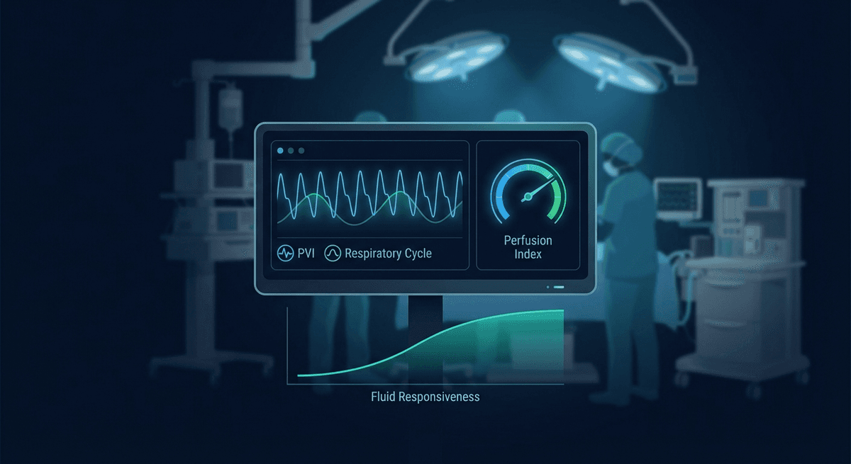
PVI = (PI_max - PI_min) / PI_max × 100%
Where PI_max and PI_min are the maximum and minimum perfusion index values over a respiratory cycle.
What PVI Tells You: Fluid Responsiveness
In a patient who is relatively hypovolemic, the right ventricle is operating on the steep portion of the Frank-Starling curve. Even small changes in preload (from intermittent positive-pressure ventilation) produce large swings in stroke volume and thus large PVI values. In a well-filled patient, the ventricle is on the plateau, and the same respiratory cycle produces minimal PVI variation.
A PVI threshold above 14% predicts fluid responsiveness (a cardiac output increase of 10% or more following a fluid bolus) with sensitivity of 81–83% and specificity of 80–84% in multiple validation studies. This makes PVI a useful guide for intraoperative fluid management, particularly in goal-directed fluid therapy protocols.
A landmark study by Cannesson et al. (2011, doi:10.1097/ALN.0b013e31820e2ab7) established PVI as a dynamic predictor of fluid responsiveness in a multicenter prospective cohort, and it is now incorporated into several institutional goal-directed therapy algorithms.
Limitations of PVI
PVI requires specific physiological conditions to be interpretable:
- Controlled mechanical ventilation: PVI is unreliable in spontaneously breathing patients because respiratory effort is irregular and generates variable pleural pressure changes
- Tidal volume dependency: PVI only accurately predicts fluid responsiveness when tidal volume is 8–10 mL/kg predicted body weight; low-tidal-volume protective ventilation (6 mL/kg) reduces PVI sensitivity
- Cardiac arrhythmias: Irregular rhythms (AF, frequent ectopics) produce PVI variation independent of fluid status
- Open chest conditions: Pericardiectomy or open thorax procedures eliminate the pleural pressure transmission mechanism
- Right heart failure: Severe right ventricular dysfunction produces chronically elevated PVI regardless of volume status
Despite these constraints, PVI remains one of the most practically accessible dynamic preload responsiveness indicators, requiring only a standard pulse oximeter — a device already present in every operating room.
Perfusion Index (PI)
Perfusion index is the ratio of the pulsatile to non-pulsatile component of the PPG signal, expressed as a percentage:
PI = (PPG_AC / PPG_DC) × 100%
Normal PI at the finger is approximately 1–20%, with higher values indicating stronger peripheral perfusion. PI below 1% at the finger indicates poor peripheral perfusion, which can result from vasoconstriction, hypovolemia, hypothermia, or low cardiac output.
Clinical Applications of PI in Perioperative Care
Peripheral vasoconstriction detection: PI drops rapidly with peripheral vasoconstriction from any cause. During neuraxial anesthesia (spinal or epidural), PI characteristically increases due to sympathetic blockade causing vasodilation. PI can be used to verify successful sympathetic block and guide vasopressor use.
Hemorrhage and hypovolemia: Decreasing PI during surgery is an early warning of hemodynamic deterioration, often preceding changes in blood pressure or heart rate. Studies by Lima et al. demonstrated that PI below 1.4% in ICU patients predicted the need for vasopressor support with reasonable accuracy.
Vasopressor effect monitoring: Following administration of vasopressors (norepinephrine, vasopressin), PI decreases as peripheral vascular resistance increases. The magnitude and duration of the PI response can help titrate vasopressor dosing.
Regional anesthesia success assessment: After nerve blocks, PI increases in the blocked region as sympathetic tone is reduced. Comparison of PI between the blocked and unblocked limb can confirm successful neural blockade.
PI and Neonatal/Pediatric Monitoring
PI has particular value in neonatal intensive care. Newborns have limited ability to mount a heart rate response to hemodynamic stress, and blood pressure measurement is technically challenging. PI provides a continuous, non-invasive window into peripheral perfusion status. PI below 0.7% in neonates correlates with increased morbidity.
A systematic review by de Graaff et al. (2019, doi:10.1097/ALN.0000000000002838) found that PI outperformed capillary refill time and other standard clinical assessments for detecting compromised perfusion in neonates and children.
PPG Waveform Analysis Beyond PVI and PI
Beyond PVI and PI, clinicians and researchers are exploring richer waveform analyses:
Respiratory rate from PPG: During controlled ventilation, the respiratory rate can be verified from the low-frequency oscillation in PPG amplitude (the same oscillation PVI exploits). During spontaneous breathing, respiratory rate estimation from PPG provides a non-invasive continuous substitute for impedance pneumography in some monitoring contexts.
PPG-based cardiac output estimation: Various algorithms attempt to estimate stroke volume from PPG waveform morphology (area under the systolic upstroke, peak-to-peak amplitude, dicrotic notch timing). These algorithms have not demonstrated sufficient accuracy for routine clinical use but remain active research areas, particularly combined with ML approaches.
Hemoglobin estimation from PPG: Multi-wavelength PPG (using 4–8 wavelengths across the visible and near-infrared spectrum) can estimate total hemoglobin non-invasively. Masimo's SpHb technology received FDA clearance for continuous hemoglobin trending, though absolute accuracy limitations (±1–2 g/dL) mean it supplements rather than replaces laboratory hemoglobin measurement.
Devices in Clinical Use
Several commercial devices provide PVI and PI monitoring:
Masimo Radical-7 and Root: The market leaders for clinical PVI/PI monitoring. The Radical-7 pulse oximeter displays PI continuously and PVI when connected to compatible probes. Masimo's Rainbow SET technology also provides SpHb and SpCO.
Nonin Cerebral/Somatic Oximetry: Not wrist-based, but regional cerebral oximetry from forehead sensors uses near-infrared spectroscopy (NIRS) principles related to PPG to monitor brain tissue oxygenation during cardiac surgery and carotid endarterectomy.
Research-grade monitors: Phillips IntelliVue and GE Healthcare monitors support advanced waveform analysis including some PVI functionality.
Integration With Goal-Directed Therapy Protocols
Modern perioperative hemodynamic management increasingly uses goal-directed therapy (GDT): using hemodynamic monitoring parameters to guide fluid and vasopressor decisions in real time rather than relying on fixed algorithms.
PVI fits well into GDT protocols as a non-invasive, continuous dynamic preload responsiveness indicator:
- Patient on controlled mechanical ventilation
- PVI > 14%: consider fluid challenge (250 mL crystalloid over 5 minutes)
- If cardiac output or blood pressure improves, continue
- If PVI normalized (<14%) and hemodynamics are stable, move to maintenance
- If PVI remains high despite adequate fluids, vasopressor therapy may be more appropriate than additional fluids
This protocol structure has been validated in abdominal surgery, orthopedic surgery, and cardiac surgery populations, with most GDT studies showing reduced intraoperative fluid use, fewer complications, and shorter hospital stays compared to fixed-rate crystalloid infusion.
Internal Links
- For perfusion index fundamentals: PPG Perfusion Index Clinical
- For PPG waveform analysis techniques: PPG Waveform Decomposition
- For respiratory monitoring from PPG: PPG Respiratory Rate Estimation
FAQ
What is the plethysmographic variability index (PVI)? PVI is the respiratory-induced variation in the PPG waveform amplitude during mechanical ventilation. It is calculated as (PI_max - PI_min) / PI_max × 100%. A PVI above 14% predicts that a patient will respond to a fluid bolus with increased cardiac output, making it a non-invasive guide for intraoperative fluid management.
How is perfusion index used in perioperative care? Perfusion index (PI) measures the ratio of pulsatile to non-pulsatile PPG signal, indicating peripheral perfusion strength. In perioperative care, it is used to detect peripheral vasoconstriction, verify neuraxial anesthesia success, monitor vasopressor response, and provide early warning of hemodynamic deterioration.
Can PPG monitor hemoglobin levels during surgery? Multi-wavelength PPG technology (Masimo SpHb) can non-invasively estimate hemoglobin continuously with accuracy within approximately 1–2 g/dL. This is insufficient to replace laboratory hemoglobin measurement but provides useful trending information to guide transfusion timing.
What are the limitations of PVI for fluid management? PVI requires controlled mechanical ventilation with adequate tidal volumes (8–10 mL/kg), regular cardiac rhythm, and absence of open-chest conditions. It is not reliable in spontaneously breathing patients, in arrhythmias, during low-tidal-volume ventilation, or with significant right ventricular dysfunction.
Is PPG-based hemodynamic monitoring replacing invasive arterial lines? PPG-based parameters are increasingly used as non-invasive complements to invasive monitoring, not direct replacements. For high-risk cardiac surgery or hemodynamically unstable patients, invasive arterial line with beat-to-beat pressure is still standard. For lower-risk procedures, PPG-based monitoring provides sufficient hemodynamic information with lower complication risk.
How accurate is PPG for estimating cardiac output? Current PPG-based cardiac output estimation algorithms have mean absolute errors of 0.5–1.5 L/min compared to thermodilution reference methods, with wide limits of agreement. This is generally insufficient for precise clinical titration but may provide useful trend information in stable patients.
Frequently Asked Questions
- What is the plethysmographic variability index (PVI)?
- PVI is the respiratory-induced variation in PPG waveform amplitude during mechanical ventilation. A PVI above 14% predicts that a patient will respond to a fluid bolus with increased cardiac output, guiding intraoperative fluid management.
- What are the limitations of PVI for fluid management?
- PVI requires controlled mechanical ventilation with adequate tidal volumes, regular cardiac rhythm, and absence of open-chest conditions. It is not reliable in spontaneously breathing patients, with arrhythmias, or during low-tidal-volume ventilation.
- How is perfusion index used in perioperative care?
- Perfusion index measures peripheral perfusion strength from the PPG signal. It detects vasoconstriction, verifies neuraxial anesthesia success, monitors vasopressor response, and provides early warning of hemodynamic deterioration.