PPG Wearable Form Factors: Ring, Patch, Earbud, and Headband Sensor Design
**The body location where a PPG sensor is worn fundamentally determines its signal quality, motion artifact susceptibility, power budget, and clinical...
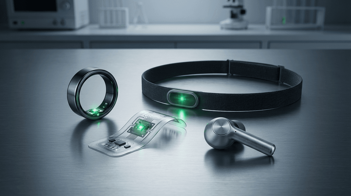
The body location where a PPG sensor is worn fundamentally determines its signal quality, motion artifact susceptibility, power budget, and clinical utility. While the underlying physics of photoplethysmography remains the same regardless of placement -- LED light enters tissue, interacts with pulsating arterial blood, and is detected by a photodiode -- the engineering constraints and physiological characteristics vary dramatically across measurement sites. This article provides a technical comparison of the five major wearable PPG form factors: wrist-worn devices, finger rings, adhesive patches, earbuds, and headbands. For each, we examine the vascular anatomy, signal characteristics, motion artifact profiles, and design trade-offs that determine measurement performance. For a foundational understanding of PPG measurement principles, see our PPG technology overview.
Vascular Anatomy and Signal Quality by Body Site
PPG signal quality is primarily determined by three anatomical factors at the measurement site: arterial blood volume fraction, epidermal thickness (which attenuates the optical signal before reaching pulsatile tissue), and proximity to large-caliber arteries versus capillary beds.
The fingertip has long been the gold standard for PPG measurement. The palmar digital arteries and their terminal branches create an exceptionally rich vascular bed, with arteriovenous anastomoses (AVAs) that produce large blood volume pulsations. The perfusion index (AC/DC ratio) at the fingertip typically ranges from 1-10%, compared to 0.2-2% at the wrist (Tamura et al., 2014; DOI: 10.3390/electronics3020282). The epidermis is approximately 0.4-0.6 mm thick on the palmar side, providing a balance between optical transmission and sensor coupling stability.
The wrist presents a more challenging environment. The radial and ulnar arteries run deep (3-5 mm below the skin surface), and the superficial vascular bed at the dorsal wrist consists primarily of venous networks and small arterioles. The dorsal skin has relatively thin epidermis (~0.1 mm) but a thicker dermis (1.5-2.5 mm) compared to the fingertip. PPG signal strength at the wrist is typically 3-10x weaker than at the fingertip, requiring higher LED drive currents and more sensitive signal processing.
The ear canal offers a surprisingly favorable measurement site. The anterior auricular artery and branches of the superficial temporal artery supply the ear canal with relatively good perfusion. The skin lining the ear canal is thin (0.5-1.0 mm), and the enclosed environment provides thermal stability that reduces vasomotor fluctuations. Budidha and Kyriacou (2018) demonstrated that ear canal PPG signals had a perfusion index of 1.5-4.5%, comparable to fingertip values and significantly higher than wrist measurements (Budidha & Kyriacou, 2018; DOI: 10.1088/1361-6579/aae7d2).
The forehead (temporal region) provides access to branches of the superficial temporal artery, yielding moderate perfusion index values of 0.5-2.0%. The thin frontal skin and minimal subcutaneous fat in the temporal region allow relatively good optical coupling. Importantly, the forehead is less susceptible to peripheral vasoconstriction during cold exposure or hemodynamic instability, making it a preferred clinical monitoring site for patients with poor peripheral perfusion.
The chest has lower pulsatile signal amplitude (perfusion index 0.1-0.8%) but offers the advantage of proximity to the heart, enabling respiratory and cardiac motion sensing through mechanical pulsation in addition to optical plethysmography.
Wrist-Worn Devices: The Dominant Consumer Form Factor
Wrist-worn PPG sensors dominate the consumer market through smartwatches and fitness trackers. Their success stems from social acceptability, large battery capacity (200-500 mAh), display integration, and established user behavior around wristwatch wearing.
Optical Design
Modern wrist PPG sensors employ multiple LEDs (typically 2-8) and multiple photodiodes (1-4) arranged in configurations optimized through Monte Carlo photon transport simulation. The Apple Watch Series 9 uses four clusters of green LEDs with a central photodiode array, operating at a primary wavelength of 525 nm for heart rate and adding red (660 nm) and infrared (940 nm) for SpO2 measurement.
Source-detector spacing of 3-5 mm is standard for green wavelengths at the wrist, balancing arterial depth penetration against signal attenuation. The optimal spacing is determined by the competing requirements of reaching pulsatile arterial tissue (which favors larger spacing) and maintaining sufficient detected photon count (which favors smaller spacing). Monte Carlo simulations show that detected signal intensity decreases approximately exponentially with spacing, with an effective attenuation coefficient of 1.5-3.0 cm^-1 depending on tissue composition (Fallow et al., 2013; DOI: 10.1117/1.JBO.18.1.017004). For more on these simulation techniques, see our photon diffusion modeling guide.
Motion Artifact Challenge
The wrist is arguably the most challenging body site for motion artifact rejection in PPG. During activities involving arm movement (walking, running, cooking, gesticulating), the sensor experiences:
- Sensor-skin displacement: Lateral and normal movement of the watch case relative to skin, changing the optical coupling geometry. Typical displacement amplitudes are 0.5-3 mm during walking.
- Tendon motion: The extensor tendons crossing the dorsal wrist slide beneath the sensor during hand flexion/extension, creating subsurface tissue displacement artifacts.
- Venous pooling: Gravity-driven blood redistribution during arm swing modulates the DC signal baseline.
- Ambient light leakage: Watch lift-off from the skin surface during motion allows ambient light to reach the photodetector.
The spectral overlap between motion artifact (0.8-3.5 Hz for walking/running cadence and harmonics) and cardiac signal (0.8-3.3 Hz for 48-200 BPM) makes simple frequency-domain separation impossible. Modern wrist devices address this through multi-axis accelerometer-referenced adaptive filtering, often combined with spectral peak tracking algorithms. Deep learning approaches trained on large motion-corrupted datasets have pushed heart rate estimation accuracy to mean absolute errors of 2-4 BPM during running, though errors can exceed 10 BPM during high-intensity interval training (Biswas et al., 2019; DOI: 10.3390/s19143299). Our motion artifact removal guide covers these algorithms in detail.
Power Consumption
Continuous green LED PPG monitoring at 25 Hz sampling typically consumes 0.5-2.0 mW, depending on LED drive current (1-25 mA), duty cycle (1-10%), and photodetector bias. Adding red and infrared channels for SpO2 increases consumption to 1.5-4.0 mW. With 300 mAh battery capacity and a 1-2 W total device power budget shared across display, processor, wireless connectivity, and sensors, continuous PPG monitoring accounts for approximately 5-15% of total power draw.
Ring-Based PPG Sensors
Ring-based PPG sensors have emerged as a compelling alternative to wrist-worn devices, offering superior signal quality from the finger's favorable vascular anatomy. The Oura Ring (generations 2 and 3), RingConn, and Ultrahuman Ring Air are leading commercial examples.
Signal Quality Advantages
The palmar digital arteries run along each side of the finger, providing a strong pulsatile blood source directly beneath a ring sensor positioned on the palmar aspect. The perfusion index from ring sensors is typically 2-8%, a 3-10x improvement over wrist measurements (de Zambotti et al., 2018; DOI: 10.3390/s18010125). This higher signal amplitude translates directly to better signal-to-noise ratio for heart rate, HRV, and SpO2 estimation.
Ring sensors also benefit from more stable skin contact. The circumferential form factor maintains consistent pressure and optical coupling around the finger, unlike a watch that can shift on the wrist. The finger has minimal subcutaneous fat between the skin surface and the arterial vessels, reducing the optical path length through non-pulsatile tissue.
Validation studies of the Oura Ring have demonstrated heart rate accuracy within 1.2 BPM mean absolute error during sleep compared to ECG reference, and HRV (RMSSD) correlation coefficients exceeding r = 0.98 against ECG-derived HRV during overnight recordings (Altini & Kinnunen, 2021; DOI: 10.3390/s21134302). For SpO2 measurement, ring sensors achieve root-mean-square errors of 1.5-2.5% compared to finger transmission pulse oximeters.
Engineering Constraints
The ring form factor imposes severe miniaturization constraints. The total volume available for electronics, battery, and sensor is approximately 0.5-1.5 cm^3, compared to 10-20 cm^3 in a smartwatch. Battery capacity is limited to 15-40 mAh, requiring aggressive power management. The Oura Ring Gen 3 achieves 4-7 day battery life by using intermittent sampling (measuring for 2-5 minutes every 5-15 minutes during the day, continuous during sleep) and low-power infrared LEDs rather than power-hungry green LEDs.
Ring size variability across users (US sizes 6-13, corresponding to 16.5-22.3 mm inner diameter) means the sensor-to-artery distance and curvature vary, affecting optical coupling. Manufacturers address this through adaptive LED drive current and multi-detector configurations that can accommodate different finger diameters.
Heat dissipation is another constraint. The small thermal mass of a ring means that even modest LED drive currents (>10 mA) can cause noticeable skin warming. LED duty cycling (on for 100-500 microseconds per sample, off between samples) limits the average thermal dissipation to below the 43 degrees C skin comfort threshold mandated by IEC 60601-1.
Motion Artifact Profile
Ring sensors have a different motion artifact profile than wrist sensors. They are relatively immune to arm swing artifacts during walking and running because the finger moves as a rigid body with the ring. However, they are highly susceptible to artifacts from hand activities: typing, gripping, lifting, and finger flexion create direct mechanical stress on the palmar digital arteries and can produce artifact amplitudes 10-100x larger than the pulsatile signal. This is why ring-based health metrics are most accurate during sleep and sedentary periods.
Adhesive Patch Sensors
Adhesive PPG patches represent the clinical research form factor, enabling continuous monitoring at body sites not served by consumer wearables. Patches are typically applied to the chest, forehead, or neck and can provide multi-day continuous recordings.
Design and Application
A typical PPG patch sensor consists of a flexible PCB (10-30 mm diameter, 2-5 mm thick) with integrated LEDs, photodiode, analog front-end, microcontroller, Bluetooth Low Energy radio, and a rechargeable or primary cell battery (50-200 mAh). The patch is affixed to the skin using medical-grade adhesive (3M 2477P or Scapa healthcare adhesives are common choices) that maintains adhesion for 1-14 days depending on the application.
The chest location provides unique measurement capabilities. While the PPG pulsatile signal amplitude is relatively low (perfusion index 0.1-0.8%), the mechanical coupling of the patch to the chest wall enables seismocardiography (SCG) through an integrated accelerometer. The combination of PPG and SCG enables pre-ejection period (PEP) estimation, which combined with pulse arrival time (PAT) provides a more accurate pulse transit time (PTT) for blood pressure estimation (Carek et al., 2017; DOI: 10.1109/TBME.2016.2606614).
Forehead patches exploit the favorable perfusion of the temporal region and its resistance to peripheral vasoconstriction. The Masimo Radius PPG forehead sensor, designed for clinical use, achieves SpO2 accuracy (Arms) of 1.5-2.0% over the 70-100% saturation range, comparable to finger probe performance and superior to wrist-based SpO2.
Advantages for Research
Patches offer several advantages for clinical research studies. The fixed position eliminates repositioning artifacts between measurement sessions. Continuous recording over 24-72 hours captures diurnal physiological variations missed by intermittent spot-check measurements. The ability to place sensors at multiple body sites simultaneously enables pulse wave velocity measurements and regional perfusion comparison studies.
The BioStamp nPoint (MC10) and Byteflies Sensor Dot are examples of research-grade flexible PPG patches that have been used in clinical studies. Patterson et al. (2020) used a forehead PPG patch to demonstrate continuous heart rate monitoring in hospitalized patients with a mean absolute error of 2.1 BPM over 48-hour recordings, with 94% of data epochs meeting a +/- 5 BPM accuracy threshold (Patterson et al., 2020).
Limitations
Adhesive patches face challenges with skin irritation (particularly in multi-day applications), adhesive failure from perspiration, and limited reusability. The rigid sensor components on flexible substrates are vulnerable to mechanical fatigue from skin stretching. Battery life constrains recording duration, with typical patches achieving 24-72 hours of continuous PPG sampling at 25-50 Hz.
In-Ear (Earbud) PPG Sensors
The ear canal has emerged as a promising site for PPG sensing, combining favorable signal characteristics with the social acceptability and market penetration of wireless earbuds. Over 300 million true wireless earbuds were sold globally in 2024, creating a massive potential platform for health sensing.
Physiological Advantages
The ear canal offers several physiological advantages for PPG. The superficial temporal artery sends branches to the external ear and ear canal, providing perfusion comparable to the fingertip. The ear canal skin is thin and well-vascularized, yielding perfusion index values of 1.5-4.5% (Budidha & Kyriacou, 2018). Crucially, the ear canal is a thermally stable, enclosed environment that minimizes ambient light interference and reduces vasomotor fluctuations caused by environmental temperature changes.
Motion artifacts in the ear canal are primarily caused by jaw movement (talking, chewing) and head rotation, which have different spectral characteristics than limb motion artifacts. Walking and running cause relatively little ear canal movement compared to the wrist, making in-ear PPG potentially more robust during lower-body exercise.
Technical Implementation
In-ear PPG sensors must fit within the tight volume constraints of an earbud. Sensor modules as small as 2 x 2 x 1 mm have been demonstrated, integrating a green or infrared LED and photodiode. The sensor is typically positioned in the concha or outer ear canal, facing the skin surface. Custom eartip designs with optical windows ensure consistent sensor-to-skin contact across different ear anatomies.
Poh et al. (2021) developed an in-ear PPG system that achieved heart rate accuracy within 1.8 BPM MAE during sedentary activities and 3.2 BPM MAE during walking, with SpO2 accuracy of 2.1% Arms compared to a finger pulse oximeter. The system used an adaptive notch filter to suppress jaw-movement artifacts and a green LED at 525 nm.
Commercial implementations include the Amazfit PowerBuds and Jabra Elite Sport, though these represent early-generation products with limited clinical validation. Apple, Samsung, and Google have filed extensive patent portfolios covering in-ear biometric sensing, suggesting more advanced implementations are forthcoming.
Unique Capabilities
In-ear PPG enables measurements that are difficult or impossible at other body sites. Core body temperature estimation from the ear canal (which reflects tympanic temperature, a close proxy for core temperature) can be combined with PPG for fever detection and thermoregulatory monitoring. EEG electrodes integrated into earbuds alongside PPG sensors enable simultaneous brain activity and cardiovascular monitoring for sleep staging, with studies showing 4-stage sleep classification accuracy of 80-85% using combined ear-EEG and PPG features (Mikkelsen et al., 2019; DOI: 10.1088/1741-2552/ab50b6).
Headband and Forehead Sensors
Headband-form PPG sensors target applications including sleep monitoring, cognitive performance tracking, meditation, and clinical monitoring of patients with poor peripheral perfusion.
Forehead PPG Characteristics
The forehead provides PPG signals through branches of the supraorbital and supratrochlear arteries (from the ophthalmic artery) and the superficial temporal artery. A key clinical advantage is that forehead perfusion is maintained during states of peripheral vasoconstriction, including hypothermia, hypovolemic shock, and use of vasoconstrictor medications. Shelley et al. (2005) demonstrated that forehead PPG maintained usable signal quality in 95% of post-cardiac-surgery patients, compared to only 59% for finger PPG, due to central-to-peripheral perfusion gradient (Shelley et al., 2005; DOI: 10.1213/01.ANE.0000155393.83818.66).
Reflectance-mode PPG on the forehead benefits from the thin skin (epidermis ~0.1 mm, dermis ~1.0 mm) and minimal subcutaneous fat in the temporal region. Source-detector spacings of 5-10 mm are optimal for the forehead, larger than the wrist due to deeper arterial targets.
Consumer and Research Implementations
The Muse S headband combines PPG with EEG for meditation and sleep tracking. The DREEM headband uses forehead PPG as part of a multi-sensor sleep staging system that achieved 85% agreement with polysomnography for 5-stage sleep classification across 25,000 nights of data (Arnal et al., 2020; DOI: 10.1038/s41591-020-0960-9). Sports headbands from Vingo and other startups target heart rate monitoring during exercise, exploiting the forehead's relative immunity to arm-motion artifacts.
Headband compliance remains a challenge for daily use, limiting this form factor primarily to sleep, exercise, and clinical applications rather than 24/7 monitoring. Hair interference with the optical path can degrade signal quality at temporal measurement sites, requiring careful sensor placement over hairless skin regions.
Comparative Performance Summary
The following summarizes typical performance across form factors, based on published validation studies:
Heart Rate Accuracy (MAE vs. ECG during rest / moderate exercise):
- Wrist: 1-3 BPM / 3-8 BPM
- Ring: 1-2 BPM / 2-4 BPM (limited exercise data)
- Patch (chest): 1-3 BPM / 2-5 BPM
- Earbud: 1-3 BPM / 3-6 BPM
- Headband: 1-2 BPM / 2-5 BPM
SpO2 Accuracy (Arms vs. co-oximetry):
- Wrist: 2.5-4.0%
- Ring: 1.5-2.5%
- Patch (forehead): 1.5-2.5%
- Earbud: 2.0-3.0%
- Headband: 1.5-2.5%
Typical Perfusion Index Range:
- Wrist: 0.2-2.0%
- Ring: 2.0-8.0%
- Patch (chest): 0.1-0.8%
- Earbud: 1.5-4.5%
- Headband: 0.5-2.0%
These values represent typical published results and vary with specific device implementation, population studied, and activity conditions. For a deeper analysis of the accuracy gap between clinical and consumer devices, see our medical vs. consumer PPG comparison.
Design Selection Guidelines
Choosing the optimal PPG form factor requires balancing competing requirements:
-
Signal quality priority (clinical accuracy, SpO2, HRV): Ring or earbud sensors provide the strongest pulsatile signals with the least motion artifact during daily activities.
-
Exercise heart rate priority: Wrist sensors with multi-LED arrays and accelerometer-based artifact rejection are mature and well-validated. Headband sensors offer a motion-resistant alternative for running.
-
Continuous clinical monitoring: Adhesive patches provide the most stable long-term recordings with minimal user interaction. Forehead patches maintain perfusion during hemodynamic instability.
-
Multi-day battery life priority: Ring sensors (4-7 days) and patches (1-3 days) outperform wrist devices (1-2 days with continuous sensing) due to lower display and processing overhead.
-
User compliance priority: Wrist devices benefit from established behavior patterns. Earbuds are increasingly accepted for daily use. Rings offer unobtrusive monitoring. Patches and headbands have lower long-term compliance.
For sensor designers and researchers, the choice of form factor is the first and most consequential decision in a PPG system design. It determines the optical configuration, power budget, motion artifact profile, and ultimately the clinical claims that can be supported. Explore the algorithmic foundations for all form factors in our algorithms reference and learn about the signal processing pipeline in our PPG technology guide.
References
- Budidha and Kyriacou. (2018). ear canal. https://doi.org/10.1088/1361-6579/aae7d2
- Shelley et al. (2005). https://doi.org/10.1213/01.ANE.0000155393.83818.66