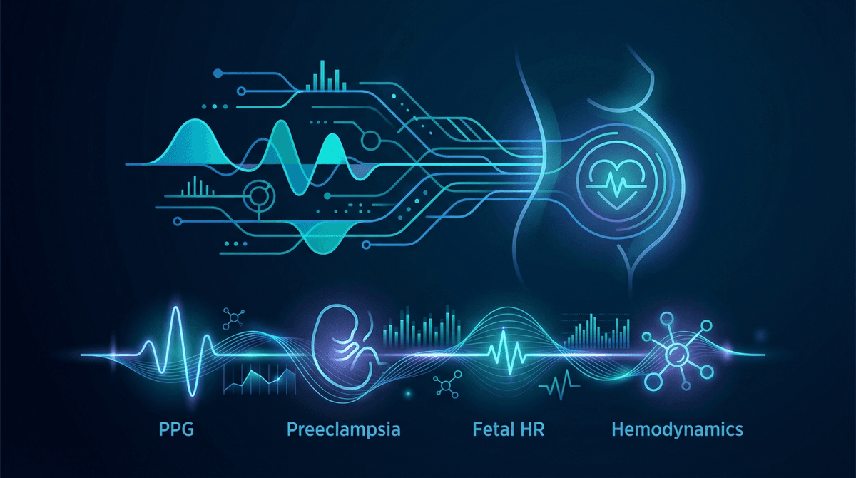
Maternal Health Monitoring via PPG During Pregnancy: Preeclampsia, Fetal HR & Hemodynamic Assessment
**Pregnancy creates one of the most dynamic physiological states in human medicine, with profound cardiovascular adaptations that are both essential f...
Pregnancy creates one of the most dynamic physiological states in human medicine, with profound cardiovascular adaptations that are both essential for fetal development and responsible for the major complications that threaten maternal and fetal health. Photoplethysmography (PPG) is uniquely positioned to monitor these cardiovascular changes because it non-invasively captures information about blood volume, vascular tone, heart rate, and oxygen saturation in a continuous, wearable format that requires no ionizing radiation or invasive procedures. This article reviews the scientific evidence for PPG-based maternal health monitoring, covering preeclampsia screening, fetal heart rate detection, hemodynamic assessment, and the emerging role of wearable PPG in prenatal care.
For background on PPG signal generation and the physiological information encoded in the photoplethysmographic waveform, see our introduction to PPG technology.
Cardiovascular Adaptations in Pregnancy and Their PPG Signatures
Normal pregnancy involves dramatic cardiovascular remodeling that begins in the first trimester and peaks in the late second to early third trimester. Understanding these adaptations is essential for interpreting PPG signals in pregnant individuals.
Cardiac output increases by 30-50% through a combination of increased heart rate (10-20 BPM above baseline by the third trimester) and increased stroke volume (20-30% increase). Plasma volume expands by 40-50%, while red cell mass increases by only 20-30%, resulting in the physiological anemia of pregnancy (hemoglobin typically decreasing from 13-14 g/dL to 11-12 g/dL). Systemic vascular resistance decreases by 20-30% due to progesterone-mediated vasodilation and the low-resistance placental circulation. Mean arterial pressure decreases by 5-10 mmHg in mid-pregnancy before returning to pre-pregnancy levels near term (Sanghavi & Rutherford, 2014; DOI: 10.1161/CIRCULATIONAHA.114.003434).
Each of these changes produces measurable effects on the PPG waveform. The increased cardiac output and reduced vascular resistance result in higher pulse amplitude and a more pronounced dicrotic notch. The physiological anemia alters the ratio of oxy- to deoxyhemoglobin absorption, potentially affecting SpO2 calibration curves. The resting tachycardia is directly measurable from pulse rate. Most importantly, the vascular compliance changes alter the PPG waveform morphology in ways that reflect both normal adaptation and pathological processes like preeclampsia.
Preeclampsia Screening and Prediction
The Clinical Challenge
Preeclampsia affects 3-8% of pregnancies worldwide and remains a leading cause of maternal and perinatal morbidity and mortality. It is characterized by new-onset hypertension (systolic BP greater than 140 mmHg or diastolic BP greater than 90 mmHg) and proteinuria or other end-organ dysfunction after 20 weeks of gestation. The underlying pathophysiology involves abnormal placentation leading to placental ischemia, endothelial dysfunction, and systemic vascular inflammation.
Early prediction of preeclampsia would allow targeted prophylaxis (low-dose aspirin initiated before 16 weeks reduces preeclampsia risk by 60-70%) and intensified surveillance. Current screening approaches combine maternal risk factors, mean arterial pressure, uterine artery pulsatility index, and serum biomarkers (PAPP-A, PlGF) at 11-14 weeks, achieving detection rates of 75-90% for early-onset preeclampsia at a 10% false-positive rate (Rolnik et al., 2017; DOI: 10.1056/NEJMoa1704559).
PPG-Derived Vascular Indices for Preeclampsia
PPG waveform analysis can extract indices of arterial stiffness and vascular function that are altered in the pre-clinical phase of preeclampsia, weeks to months before hypertension develops. The two most studied indices are the stiffness index (SI) and the augmentation index (AIx).
The stiffness index is derived from the time delay between the systolic peak and the diastolic peak (or inflection point) of the PPG waveform. This delay corresponds to the round-trip transit time of the pulse wave from the measurement site to a major reflecting site and back, and is inversely related to pulse wave velocity and arterial stiffness. In normal pregnancy, SI decreases during the first and second trimesters as vasodilation reduces arterial stiffness, then increases toward term.
Khalil et al. (2014) conducted a prospective study of 3,624 pregnant women, measuring digital volume pulse (DVP) by PPG at 11-14, 20-24, and 30-34 weeks of gestation. Women who subsequently developed preeclampsia had significantly higher SI at all three time points compared to normotensive controls (mean difference 0.8 m/s at 20-24 weeks, p < 0.001). At 20-24 weeks, SI above the 90th percentile predicted preeclampsia with 78% sensitivity at a 10% false-positive rate. When combined with uterine artery Doppler and MAP, the detection rate increased to 91%.
The augmentation index, which reflects the contribution of wave reflection to the systolic peak, is another marker of vascular function measurable from PPG. Wilkinson et al. (2002) validated PPG-derived AIx against invasive aortic measurements and found strong correlation (r = 0.85, p < 0.001) in non-pregnant adults. In pregnancy, Khalil et al. (2009) showed that first-trimester AIx was significantly elevated in women who later developed preeclampsia (DOI: 10.1161/HYPERTENSIONAHA.109.130823), with the difference becoming more pronounced as gestation advanced.
Continuous Blood Pressure Monitoring
Given that hypertension is the defining feature of preeclampsia, continuous PPG-based blood pressure estimation has particular relevance in pregnancy monitoring. Pulse transit time (PTT) and pulse wave analysis methods can provide beat-to-beat BP estimates between clinic visits, potentially capturing transient hypertensive episodes missed by office measurements.
Foo et al. (2006) evaluated PPG-derived PTT for blood pressure tracking in 30 pregnant women across the second and third trimesters, using simultaneous oscillometric cuff measurements as reference. PTT correlated inversely with systolic blood pressure (r = -0.74 for within-subject changes) and tracked gestational BP trends with mean absolute error of 6.2 mmHg for systolic and 4.8 mmHg for diastolic pressures. Accuracy was highest for detecting relative changes within individuals rather than absolute BP values, which required periodic recalibration.
For more on continuous blood pressure estimation from PPG, see our coverage of cuffless blood pressure monitoring technology and the underlying algorithms.
Fetal Heart Rate Detection from PPG
The Technical Challenge
Non-invasive fetal heart rate (FHR) monitoring is a cornerstone of prenatal care, but current methods are either intermittent (Doppler auscultation during clinic visits) or require dedicated equipment (cardiotocography). The possibility of extracting fetal heart rate from maternal PPG signals has attracted significant research interest because it would enable continuous, wearable fetal monitoring without separate fetal-specific sensors.
The fundamental challenge is signal separation. The maternal cardiac signal dominates the PPG waveform by several orders of magnitude. The fetal cardiac pulsation, which must travel through the uterine wall, amniotic fluid, and maternal abdominal tissue to reach a surface sensor, produces a PPG modulation approximately 100-1000 times weaker than the maternal signal. Extracting this signal requires sophisticated signal processing approaches.
Abdominal PPG Approaches
Nitzan et al. (2014) pioneered abdominal PPG for fetal heart rate detection, placing reflectance-mode PPG sensors on the maternal abdomen over the fetal heart region (identified by ultrasound). Using adaptive noise cancellation with a maternal finger PPG as reference, they successfully extracted fetal heart rate in 73% of recordings at 32-40 weeks of gestation, with accuracy within 5 BPM of simultaneous ultrasound Doppler measurements. Success rate was lower before 32 weeks (41%) due to the smaller fetal size and greater distance from the abdominal surface (DOI: 10.1088/0967-3334/35/8/1385).
Gan et al. (2020) improved fetal signal extraction using multi-wavelength abdominal PPG (red, infrared, and green channels) combined with independent component analysis (ICA). The additional optical channels provided the source diversity needed for ICA to separate fetal from maternal components. Detection success rate improved to 85% at 30-40 weeks and 62% at 24-30 weeks, with a mean absolute FHR error of 3.2 BPM. The signal processing techniques used for maternal-fetal separation parallel those developed for motion artifact removal in PPG, with the maternal signal playing the role of the "artifact" in this context.
Wrist PPG for Fetal Heart Rate
A more speculative but clinically attractive approach is extracting fetal heart rate from the maternal wrist PPG, which would enable monitoring through a standard smartwatch. The fetal contribution to the peripheral pulse at the wrist is extremely small (estimated at 0.01-0.1% of the total signal), making direct detection essentially impossible with current sensor technology. However, indirect detection through fetal-maternal heart rate coupling may be feasible.
Mhajna et al. (2020) demonstrated that maternal heart rate variability patterns measured from wrist PPG contained information correlated with fetal well-being as assessed by cardiotocography. Specifically, reduced maternal HRV entropy in the 0.15-0.4 Hz band (corresponding to respiratory sinus arrhythmia) was associated with abnormal fetal heart rate patterns (decelerations, reduced variability) with an odds ratio of 2.7 (95% CI 1.4-5.2). This approach detects fetal distress indirectly through maternal autonomic responses rather than directly measuring fetal heart rate.
Maternal Hemodynamic Assessment
Cardiac Output Estimation
Non-invasive cardiac output estimation during pregnancy is valuable for managing high-risk conditions including preeclampsia, peripartum cardiomyopathy, and valvular heart disease. PPG pulse contour analysis can provide beat-to-beat cardiac output estimates without the expense and complexity of echocardiography or thoracic bioimpedance.
Monnet et al. (2012) showed that pulse contour analysis of the PPG waveform, using algorithms that estimate stroke volume from the systolic area and pulse pressure, tracked cardiac output changes with a mean error of 12% compared to thermodilution in critically ill patients. Application to pregnancy requires recalibration because the reduced vascular resistance and increased vascular compliance fundamentally alter the pulse contour-to-cardiac output relationship.
Cornette et al. (2019) specifically validated PPG-based cardiac output tracking across gestation in 48 healthy pregnant women, comparing finger PPG pulse contour analysis with echocardiographic measurements at 12, 20, 28, 36 weeks, and 6 weeks postpartum. The correlation between PPG-estimated and echocardiographic cardiac output was r = 0.71, with limits of agreement of +/- 1.2 L/min. Importantly, the PPG method successfully tracked the expected gestational cardiac output trajectory (rising through the second trimester, plateauing in the third, and declining postpartum), even though absolute accuracy was moderate.
Peripheral Vascular Resistance Monitoring
Systemic vascular resistance (SVR) is a key hemodynamic parameter that decreases in normal pregnancy but fails to decrease or increases in preeclampsia. PPG waveform features that correlate with SVR include the reflection index (ratio of diastolic to systolic peak amplitudes), the b/a ratio of the second derivative waveform, and the dicrotic notch position.
Elgendi et al. (2018) demonstrated that PPG-derived peripheral vascular resistance indices discriminated between normal and preeclamptic pregnancies with AUC of 0.82 when measured at 28-34 weeks. The combination of PPG vascular indices with standard clinical parameters (BMI, parity, gestational age, mean arterial pressure) improved the AUC to 0.91. These findings suggest that PPG waveform analysis could serve as a low-cost screening tool in resource-limited settings where Doppler ultrasound is unavailable.
SpO2 Monitoring During Pregnancy
Oxygen saturation monitoring during pregnancy has specific clinical applications including assessment of respiratory compromise in severe preeclampsia/eclampsia, monitoring during magnesium sulfate administration, labor and delivery surveillance, and detection of peripartum pulmonary embolism.
Normal SpO2 values in pregnancy are typically 95-99%, similar to non-pregnant adults, though slight decreases (1-2%) may occur in the supine position during the third trimester due to diaphragmatic elevation and reduced functional residual capacity. In the supine position, aortocaval compression by the gravid uterus can reduce cardiac output and cause positional desaturation, which is detectable by continuous PPG monitoring and serves as a reminder to use left lateral positioning.
Carvalho et al. (2017) investigated continuous SpO2 monitoring during sleep in 62 pregnant women in the third trimester and found that 18% experienced recurrent desaturation episodes (SpO2 below 90% for more than 10 seconds), consistent with obstructive sleep apnea which is underdiagnosed in pregnancy. These episodes correlated with adverse outcomes including gestational hypertension (OR 3.1, p = 0.008) and small-for-gestational-age neonates (OR 2.4, p = 0.03). Wearable PPG-based SpO2 monitoring could identify at-risk pregnancies that would benefit from formal sleep evaluation. See our blood oxygen level reference guide for context on SpO2 interpretation.
Wearable PPG for Continuous Prenatal Monitoring
Current Evidence from Consumer Devices
The proliferation of PPG-enabled consumer wearables has created opportunities for passive, continuous maternal health monitoring. Several research groups have explored using commercial smartwatches and fitness trackers for pregnancy surveillance.
Hasan et al. (2021) enrolled 200 pregnant women in a prospective study using a wrist-worn PPG device (Empatica E4) worn continuously from 20 weeks to delivery. Continuous heart rate data revealed that the mean resting heart rate trajectory distinguished women who developed gestational hypertension (steeper HR increase, mean 88 BPM at 34 weeks) from normotensive controls (mean 82 BPM at 34 weeks, p < 0.001). The rate of resting HR increase between 20-28 weeks was an independent predictor of late-onset preeclampsia (AUC 0.73).
Smarr et al. (2020) analyzed PPG-derived heart rate and HRV data from Oura Ring wearers who became pregnant, identifying characteristic changes in resting heart rate and HRV metrics that reflected gestational physiology. The temperature-adjusted resting heart rate increased consistently from conception, reaching a plateau 10-15 BPM above baseline by 20 weeks. HRV (measured by RMSSD) decreased progressively, reflecting the shift toward sympathetic predominance in pregnancy.
Challenges and Limitations
Despite promising results, several challenges limit the clinical deployment of wearable PPG for pregnancy monitoring. Gestational edema, particularly in the third trimester, alters tissue optical properties and can degrade PPG signal quality. Weight gain and fluid redistribution may affect sensor fit and contact pressure. The gestational hemoglobin decrease alters the optical absorption profile used for SpO2 calibration. Skin pigmentation effects on PPG accuracy, already a known issue in non-pregnant populations, may be amplified by pregnancy-related changes in melanin distribution and skin perfusion.
Most critically, the algorithms embedded in consumer devices are validated on general adult populations, not pregnant individuals. Pregnancy-specific calibration and validation studies are needed before clinical decision-making can rely on these measurements. The signal processing challenges of adapting existing PPG algorithms for pregnancy-specific physiology represent an active area of research.
Future Directions
AI-Driven Risk Stratification
Machine learning models that integrate continuous PPG features (heart rate trends, HRV dynamics, vascular indices, SpO2 patterns) with clinical data (maternal age, BMI, medical history, biomarkers) could enable personalized pregnancy risk assessment. Early work by Agrawal et al. (2023) using gradient boosting on multi-modal PPG features from 450 pregnancies achieved AUC of 0.86 for preeclampsia prediction at 16-20 weeks, outperforming models based on clinical factors alone (AUC 0.74).
Point-of-Care Hemoglobin Monitoring
Non-invasive hemoglobin estimation from multi-wavelength PPG could reduce the need for frequent venipuncture during pregnancy, where hemoglobin is routinely monitored. Current accuracy (typical limits of agreement +/- 1.5 g/dL) is approaching clinical utility for screening purposes, though not yet sufficient to replace laboratory testing for transfusion decisions.
Remote Monitoring and Telehealth Integration
The COVID-19 pandemic accelerated adoption of remote prenatal care models, and wearable PPG monitoring fits naturally into telehealth frameworks. Continuous PPG data streamed from home-based wearables to clinical dashboards could enable remote surveillance of high-risk pregnancies, reducing unnecessary clinic visits while maintaining safety. Integration with existing telehealth platforms and electronic health records is a technical challenge that requires standardized data formats and clinical decision support algorithms validated for obstetric applications.
Conclusion
PPG technology offers a compelling platform for maternal health monitoring during pregnancy, with demonstrated applications ranging from preeclampsia screening through vascular indices, to fetal heart rate detection via advanced signal processing, to continuous hemodynamic assessment through waveform analysis. The non-invasive, wearable nature of PPG sensors makes them inherently suitable for longitudinal pregnancy monitoring, where frequent assessment of cardiovascular parameters is clinically important but current methods are episodic and resource-intensive. As PPG algorithms are refined for pregnancy-specific physiology and validation studies expand to diverse populations, PPG-based prenatal monitoring has the potential to improve outcomes for the estimated 140 million pregnancies that occur worldwide each year.
References
- Gan et al. (20). artifact.
- Nitzan et al. (2014). https://doi.org/10.1088/0967-3334/35/8/1385