What Is Photoplethysmography? A Complete Technical Overview for 2026
Photoplethysmography (PPG) is an optical technique for measuring blood volume changes in tissue. The principle is simple: shine light into tissue, mea...
Photoplethysmography (PPG) is an optical technique for measuring blood volume changes in tissue. The principle is simple: shine light into tissue, measure how much bounces back (or passes through), and track changes in absorption caused by blood volume fluctuations with each heartbeat. The technology behind that simple principle has evolved from bulky clinical devices in the 1930s to the sensors packed into every mainstream smartwatch in 2026 — and the clinical value extracted from those signals continues to grow.
The Core Principle: Light and Blood
Blood contains hemoglobin — a protein that absorbs light. Oxygenated hemoglobin (HbO2) and deoxygenated hemoglobin (Hb) have different absorption spectra, which is why blood changes color between arteries and veins. When light enters tissue, it is absorbed and scattered. The more blood present (higher vessel volume), the more light is absorbed and the less is reflected back to a detector.
With each heartbeat, arterial blood volume in the microvessels of the tissue expands and contracts. This pulsatile volume change produces a pulsatile change in light absorption — the AC (alternating current) component of the PPG signal. Superimposed on this is a stable baseline absorption from non-pulsatile tissue components (bones, muscle, venous blood, fat) — the DC (direct current) component.
The ratio of AC to DC (the perfusion index) reflects the relative pulsatile signal strength — how much of the total optical path is dominated by arterial pulsations.
PPG Hardware: What's Actually in the Sensor
A PPG sensor has three primary components:
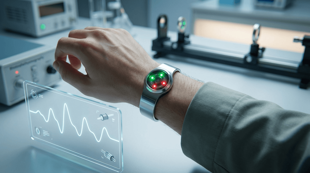
Light source (LED): One or more LEDs illuminate the tissue. LED wavelength selection determines which aspects of blood composition are measured:
- Green (530-550 nm): Strong hemoglobin absorption; excellent signal quality on dark skin; best for heart rate. Used by most wrist wearables (Apple Watch, Fitbit, Garmin).
- Red (620-700 nm): Used in pulse oximetry (SpO2) paired with infrared. Lower absorption than green.
- Infrared (800-1000 nm): Deep tissue penetration; used for SpO2, PTT measurements, and research. Less affected by melanin variation.
- Multi-wavelength: High-end devices use 4-8 LEDs at different wavelengths simultaneously, enabling more complex hemodynamic decomposition.
Photodetector: A photodiode or phototransistor measures the returned light intensity. Positioned next to the LED(s) in reflectance mode, or opposite the LED in transmission mode. Spectral sensitivity should match the LED emission wavelength.
Optical isolation: Physical separation between LED and detector, plus optical baffles, prevents LED light from directly reaching the detector without passing through tissue (crosstalk elimination).
Analog front-end (AFE): The raw photodetector signal is in the nanoampere range. An AFE IC provides transimpedance amplification, programmable gain, analog bandpass filtering, and analog-to-digital conversion. Major AFE manufacturers: Texas Instruments (AFE4900 series), Maxim Integrated (MAX86141), Analog Devices.
Signal Acquisition: Modes and Configurations
Transmission mode: LED and detector on opposite sides of tissue (finger clip, earlobe clip). Light passes through the tissue. Highest SNR; best for clinical pulse oximetry. Impractical for wrist-form-factor wearables.
Reflectance mode: LED and detector on the same surface. Light enters tissue, scatters, and returns to the detector. Lower SNR than transmission, but wearable-compatible. Standard configuration for smartwatch sensors.
Sampling rate: Clinical PPG (hospital monitors): 250 Hz standard. Consumer wearables: 25-100 Hz typical, with burst modes up to 200 Hz for research features. For accurate HRV (beat-to-beat timing), 100+ Hz is recommended to achieve sub-10ms IBI resolution.
Ambient light cancellation: LEDs are strobed (turned on/off rapidly) while the detector runs continuously. Subtraction of the OFF-state reading (ambient only) from the ON-state (LED + ambient) removes ambient light interference. Modern AFEs implement this in hardware.
The PPG Signal: AC and DC Components
The PPG signal is typically displayed after analog and digital processing that emphasizes the AC component:
DC component (low frequency, <0.5 Hz): Reflects slow changes in tissue blood volume — breathing (respiratory modulation), thermoregulation, autonomic vasomotor waves (Mayer waves at ~0.1 Hz). The DC level itself encodes perfusion state.
AC component (~0.5-5 Hz): The cardiac pulsatile component. One cycle per heartbeat. This is the primary component used for heart rate, HRV, oxygen saturation, pulse waveform analysis, and most clinical applications.
High-frequency noise (>5 Hz): Electrical noise, motion artifacts, optical interference. Removed by filtering before signal processing.
What Can PPG Measure?
The range of physiological parameters extractable from PPG has expanded dramatically as signal processing and machine learning capabilities have grown:
Heart rate: Fundamental application. Accurate to ±2 bpm at rest with modern algorithms. Degrades with motion. Applied in all consumer wearables for decades.
Heart rate variability (HRV): Beat-to-beat IBI sequence extracted from pulse peak or foot timing. Reflects autonomic nervous system state. Accurate at rest (see HRV accuracy guide), degrades with movement.
Oxygen saturation (SpO2): Ratio method comparing red and infrared AC/DC components. Validated for clinical screening; medical-grade devices require calibration to CO-oximetry.
Blood pressure: PTT-based cuffless BP estimation using two-site PPG or PPG+ECG. Research active; consumer accuracy not yet at clinical standard. See PTT blood pressure guide.
Arterial stiffness indices: PWV, stiffness index, SDPPG aging index — derived from waveform morphology. See arterial stiffness guide.
Respiratory rate: PPG amplitude, baseline, and frequency are all modulated by respiration. Multiple algorithms extract respiratory rate from single-site PPG with errors of 1-3 breaths/min at rest. See respiratory rate estimation.
Atrial fibrillation detection: Irregular IBI patterns characteristic of AF are detectable with high sensitivity and specificity using trained algorithms. FDA-cleared applications exist (Apple Watch, KardiaMobile).
Sleep staging: Overnight PPG HRV patterns, combined with accelerometry and respiratory rate, enable automated sleep stage classification. Performance approaches clinical actigraphy.
Stress detection: Sympathetic activation signatures in PPG amplitude, HRV, and waveform morphology are used in commercial stress tracking algorithms.
Pulse transit time and perfusion: Regional perfusion assessment, peripheral vascular disease screening, and microcirculation evaluation are active clinical research applications.
PPG in 2026: State of the Technology
Consumer wearable PPG has reached saturation for basic vital signs (heart rate, SpO2). The current frontier is three-dimensional:
1. Clinical-grade accuracy: Moving from consumer convenience to medical diagnostic capability requires rigorous validation, cleared regulatory pathways (FDA De Novo, CE IVDR), and robust real-world performance. Several companies are pursuing this for AF detection, hypertension screening, and sleep disorders.
2. Multi-wavelength and multimodal sensing: Combining 4-8 wavelength PPG with EDA (electrodermal activity), skin temperature, accelerometry, and barometric pressure enables richer physiological state reconstruction. Continuous hemoglobin, total hemoglobin, and tissue oxidation measurement are emerging capabilities.
3. AI-native signal processing: Deep learning models trained on millions of device-hours of labeled PPG data extract features beyond what any hand-crafted algorithm can identify. Foundation models for waveform understanding are being developed at scale.
The ChatPPG platform is designed around this convergence — providing both the signal processing toolkit for researchers and the clinical interpretation layer for understanding what the numbers mean.
PPG vs. Other Cardiovascular Monitoring Modalities
| Modality | What It Measures | Invasive? | Continuous? | Wearable? |
|---|---|---|---|---|
| PPG | Volume changes, optically | No | Yes | Yes |
| ECG | Cardiac electrical activity | No | Yes | Yes (chest) |
| Arterial line | Invasive blood pressure | Yes | Yes | No |
| Tonometry | Pressure wave (skin surface) | No | Yes | Limited |
| Echocardiography | Cardiac structure/function | No | Short sessions | No |
| cfPWV | Aortic stiffness | No | Single measurement | No |
PPG occupies a unique niche: non-invasive, continuous, wearable, and capable of encoding multiple hemodynamic parameters in a single measurement. Its limitation is the indirect nature of optical blood volume measurement — PPG is always an inference about cardiovascular state, not a direct measurement of it.
Frequently Asked Questions
What does photoplethysmography measure? Photoplethysmography measures changes in blood volume in peripheral tissue using light. It produces a pulsatile signal synchronized to each heartbeat, from which heart rate, HRV, oxygen saturation, arterial stiffness, respiratory rate, and other cardiovascular parameters can be derived.
How does a PPG sensor work? An LED shines light into tissue; a photodetector measures returning light intensity. Blood absorbs more light when vessel volume increases (systole) and less when it decreases (diastole), producing a pulsatile optical signal that tracks each heartbeat.
What is the difference between PPG and pulse oximetry? Pulse oximetry is a specific PPG application that uses two wavelengths (red and infrared) to calculate the ratio of oxygenated to total hemoglobin (SpO2). Regular PPG can use a single wavelength. All pulse oximeters use PPG; not all PPG systems measure SpO2.
What wavelength does a PPG sensor use? It depends on application. Green light (530-550 nm) is most common for heart rate measurement in wrist wearables due to strong hemoglobin absorption and resistance to melanin interference. Red (660 nm) and infrared (940 nm) are used for SpO2. Near-infrared (850+ nm) is used for deep tissue applications.
Is PPG accurate for heart rate measurement? At rest, modern PPG-based heart rate is accurate to within ±2-3 bpm compared to ECG in most conditions. Accuracy degrades with vigorous exercise and motion artifacts. Most consumer wearables use accelerometer-based motion artifact rejection to maintain accuracy during activity.
What is the difference between wrist PPG and finger PPG? Finger PPG (transmission mode, fingertip clip) has higher SNR and better waveform quality — preferred for clinical research, pulse oximetry, and detailed waveform analysis. Wrist PPG (reflectance mode) trades signal quality for convenience and continuous wearability. Wrist devices use more sophisticated algorithms to compensate.
{
  "@context": "https://schema.org",
  "@type": "FAQPage",
  "mainEntity": [
    {
      "@type": "Question",
      "name": "What does photoplethysmography measure?",
      "acceptedAnswer": {
        "@type": "Answer",
        "text": "Photoplethysmography measures changes in blood volume in peripheral tissue using light. It produces a pulsatile signal synchronized to each heartbeat, from which heart rate, HRV, oxygen saturation, arterial stiffness, and other cardiovascular parameters can be derived."
      }
    },
    {
      "@type": "Question",
      "name": "How does a PPG sensor work?",
      "acceptedAnswer": {
        "@type": "Answer",
        "text": "An LED shines light into tissue; a photodetector measures returning light intensity. Blood absorbs more light when vessel volume increases during systole and less during diastole, producing a pulsatile optical signal tracking each heartbeat."
      }
    },
    {
      "@type": "Question",
      "name": "What is the difference between PPG and pulse oximetry?",
      "acceptedAnswer": {
        "@type": "Answer",
        "text": "Pulse oximetry is a specific PPG application using two wavelengths (red and infrared) to calculate SpO2. All pulse oximeters use PPG; not all PPG systems measure SpO2."
      }
    },
    {
      "@type": "Question",
      "name": "Is PPG accurate for heart rate measurement?",
      "acceptedAnswer": {
        "@type": "Answer",
        "text": "At rest, modern PPG-based heart rate is accurate to within ±2-3 bpm compared to ECG. Accuracy degrades with vigorous exercise and motion artifacts."
      }
    },
    {
      "@type": "Question",
      "name": "What wavelength does a PPG sensor use?",
      "acceptedAnswer": {
        "@type": "Answer",
        "text": "Green light (530-550 nm) is most common for heart rate in wrist wearables. Red (660 nm) and infrared (940 nm) are used for SpO2. Near-infrared (850+ nm) is used for deep tissue applications."
      }
    }
  ]
}
{
  "@context": "https://schema.org",
  "@type": "Article",
  "headline": "What Is Photoplethysmography? A Complete Technical Overview for 2026",
  "description": "Complete technical overview of photoplethysmography (PPG): how the sensor works, what it measures, signal components, and the full range of clinical and wearable applications.",
  "author": {"@type": "Organization", "name": "ChatPPG Research Team"},
  "publisher": {"@type": "Organization", "name": "ChatPPG", "url": "https://chatppg.com"},
  "datePublished": "2026-03-21",
  "url": "https://chatppg.com/blog/ppg-photoplethysmography-sensor-overview"
}
Related reading: PPG Sensor Design Guide | LED Wavelength Selection | PPG AC/DC Ratio Explained | Start learning PPG