Neonatal Perfusion Index Explained: How PPG Supports NICU Monitoring
Understand neonatal perfusion index, how PPG derives it from the waveform, and how NICU teams use bedside trends to assess peripheral flow in newborns.
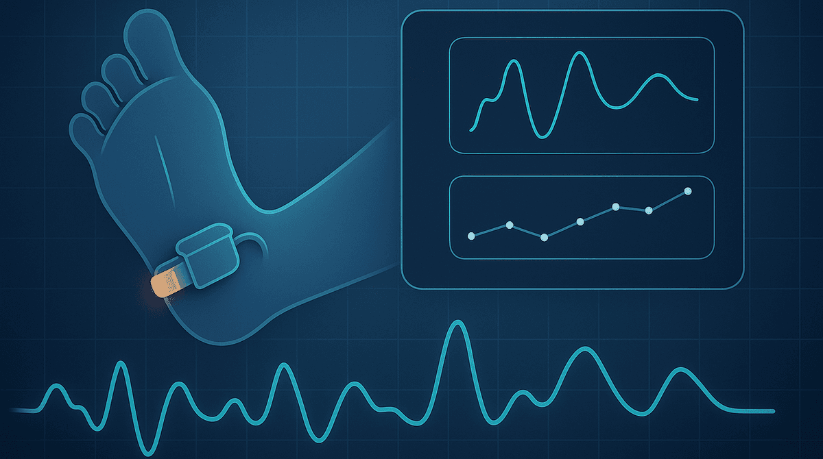
Neonatal perfusion index is a bedside estimate of how strong pulsatile blood flow is at the sensor site, derived from the photoplethysmography waveform rather than from oxygen saturation alone. In the NICU, PPG lets teams follow peripheral perfusion continuously, so changes in circulation, temperature, vascular tone, or response to treatment appear as trends instead of isolated guesses.
What neonatal perfusion index actually measures
Perfusion index, often shortened to PI, is a number calculated from the PPG signal collected by a pulse oximeter or related optical sensor. The sensor shines light through or onto tissue and measures how much light is absorbed. Part of that signal changes with each heartbeat because arterial blood volume rises and falls. Another part stays more constant because it comes from tissue, venous blood, bone, and other nonpulsatile components.
PI expresses the relationship between those two parts of the signal. In simple terms, it asks: how large is the pulsatile waveform compared with the steady background? A stronger pulse waveform at the probe site usually produces a higher PI, while a faint waveform produces a lower PI. That is why PI is best understood as a waveform-derived bedside metric, not just a side number next to SpO2.
This distinction matters in the NICU. Oxygen saturation tells you how much hemoglobin is carrying oxygen. PI tells you something different: how well the arterial pulse is showing up in the monitored tissue at that moment. A baby can have an acceptable SpO2 and still show a weak peripheral pulse waveform. The reverse can also be true.
PI is therefore not a direct measure of cardiac output, blood pressure, or organ perfusion. It is a local optical estimate of pulsatile flow at the sensor location. Used carefully, it gives clinicians another window into neonatal hemodynamics, especially when trend behavior lines up with the rest of the bedside picture.
How PPG turns a waveform into a perfusion index
Photoplethysmography works by capturing tiny blood volume changes in the microvascular bed. Every heartbeat creates an alternating component in the optical signal. This is the pulse waveform that clinicians see on the monitor. The baseline beneath it is the nonpulsatile component.
Most PI displays come from the ratio of the pulsatile component to the nonpulsatile component, often expressed as a percentage. The exact implementation can vary by device, filtering approach, and sensor design, but the bedside meaning is similar across systems. A taller, cleaner pulse waveform relative to the background produces a higher PI. A flatter waveform, or one degraded by vasoconstriction, poor contact, motion, or low local flow, produces a lower PI.
That is why the waveform should be inspected whenever PI looks odd. If the pleth trace is noisy, clipped, inconsistent, or poorly synchronized with the heart rate, the displayed number may not reflect a real physiologic shift. In neonatal care, where infants are small, active, temperature sensitive, and often monitored on hands or feet with tiny vessels, signal quality is part of interpretation, not a separate issue.
Another useful way to think about PI is that it compresses waveform amplitude into a single trendable output. Instead of relying only on visual inspection of the pleth trace, the monitor gives a numeric summary of how prominent the pulse signal is over time. That makes PI attractive for continuous bedside use, as long as clinicians remember where the number comes from.
Why perfusion index is especially relevant in the NICU
Neonates have rapidly changing circulation in the first hours and days after birth. Peripheral tone can shift with temperature, stress, respiratory support, ductal shunting, infection, and medication exposure. Preterm infants may show even greater variability because their vascular regulation is immature and their skin and tissue characteristics affect optical monitoring.
In this setting, a waveform-derived measure can be helpful because it reacts quickly. A foot probe may show the pulse becoming smaller before anyone would describe the infant as looking gray or cool. A PI trend may improve after warming, fluid adjustment, or a change in support, even before a full set of labs is back. That does not make PI a standalone diagnostic tool, but it can make the bedside team more responsive to developing patterns.
PI can also add nuance when oxygenation alone is not enough. Two infants may both have acceptable saturation values, yet one has a robust peripheral waveform and the other has a weak one. When clinicians already use PPG for heart rate and SpO2, PI offers another signal from the same optical source. For broader context on how these measures fit together, see PPG neonatal monitoring, neonatal oxygen monitoring, and continuous SpO2 monitoring with wearables.
Clinical situations where PI can be useful
1. Transition after birth
The immediate postnatal period is a time of major circulatory change. Pulmonary vascular resistance falls, shunts change direction, and peripheral perfusion can be uneven while the infant adapts. During this window, PI may help clinicians watch how strongly the peripheral pulse is being transmitted to the sensor site. A rising, stabilizing PI trend can support the impression that circulation is settling, while persistently weak or unstable values may prompt a closer look at temperature, sensor placement, respiratory status, or hemodynamics.
2. Suspected low peripheral perfusion
When a newborn is cool, pale, mottled, or slow to reperfuse, PI can serve as a continuous companion to hands-on assessment. A low PI does not diagnose shock, sepsis, or low cardiac output on its own, but it can reinforce concern when paired with other findings such as prolonged capillary refill, metabolic acidosis, weak pulses, or falling urine output. It may also help the team recognize that the pulse waveform is poor even when saturation remains measurable.
3. Tracking response to interventions
PI is often most useful when something changes and the team wants to know if the pulse waveform follows. Examples include repositioning a sensor, warming an infant, adjusting respiratory support, starting or titrating vasoactive therapy, giving fluid, or reducing agitation. If PI rises along with improved waveform quality and better overall clinical appearance, that trend can support the bedside impression that peripheral perfusion has improved.
How to interpret neonatal PI without overreading it
The biggest mistake is treating PI like a universal cutoff with the same meaning in every baby. That is rarely how it works.
A more reliable bedside approach looks like this:
- Trend first. A stable infant with a consistently moderate PI is different from an infant whose PI is falling over 20 to 30 minutes.
- Check the waveform. If the pleth trace is poor, the number may be poor for technical reasons.
- Interpret PI with other signals. Heart rate, SpO2, temperature, blood pressure, capillary refill, lactate, urine output, and clinical exam all matter.
- Know the sensor site. Hand and foot values can differ, and edema or local compression can change the reading.
- Expect context to matter. Crying, handling, cold stress, vasoconstriction, and medications can all move PI.
- Respect device differences. A threshold reported in one study or monitor family may not transfer neatly to another.
This is why many NICU teams get more value from PI as a trend line than as a single alarm threshold. The number becomes much more informative when viewed across time, against the waveform, and in light of the infant's overall status.
Common reasons PI changes in newborns
A falling PI can reflect true reduction in peripheral pulsatile flow, but several bedside factors can create the same appearance. Cold extremities are a common example. Peripheral vasoconstriction makes the pulse waveform smaller at the foot or hand even if central circulation is still being maintained. Motion artifact can do the same by corrupting the optical signal. Loose wraps, excessive ambient light, pressure on the sensor, and poor skin contact are also frequent causes.
Clinical factors matter too. Hypovolemia, sepsis, hemodynamic instability, pain, agitation, vasoactive medications, and the transition from fetal to neonatal circulation can all influence vascular tone and pulse amplitude. Preterm infants may show wider variability because their vessels and skin properties respond differently from those of term infants.
A rising PI also needs context. It may reflect improved peripheral perfusion, better probe contact, warming, or a calmer infant.
A practical bedside workflow for NICU teams
When PI is used well, it fits into normal observation rather than creating a separate monitoring silo.
- Confirm signal quality first. Look at the pleth waveform, heart rate match, and sensor position.
- Note the site and timing. Hand versus foot, preductal versus postductal context, and age after birth all shape interpretation.
- Compare PI with the clinical picture. Does the infant look warm, pink, and stable, or cool and poorly perfused?
- Trend rather than react instantly. A sustained change carries more meaning than one brief dip during handling.
- Escalate when multiple findings align. Persistent low PI plus concerning exam findings deserves more attention than PI alone.
This approach keeps PI in its proper role. It is not a replacement for examination, echocardiography, blood pressure monitoring, or laboratory assessment. It is a continuous, low-burden signal that can make the PPG waveform more actionable at the bedside.
The main takeaway
In neonatal care, perfusion index is most helpful when clinicians remember what it really represents: the relative strength of the pulsatile PPG waveform at the sensor site. That makes it a practical bedside marker of peripheral pulse signal quality and perfusion trend, especially in the NICU where circulation can change quickly.
Used with waveform review and clinical context, PI can highlight early shifts that SpO2 alone may miss. Used without that context, it can be overinterpreted. The best use of neonatal PI is thoughtful, trend-based, and tied to the physiology behind the waveform.
FAQ
What is a normal neonatal perfusion index?
There is no single normal PI value that applies to every newborn, device, and monitoring site. In practice, trend behavior and correlation with the bedside assessment are usually more useful than one fixed cutoff.
Is perfusion index the same as oxygen saturation?
No. SpO2 estimates how much hemoglobin is carrying oxygen, while PI estimates how strong the pulsatile optical signal is at the sensor site. A baby can have normal or near normal saturation and still show a low PI if the peripheral pulse waveform is weak.
Why can PI be low even when the baby looks stable?
A low PI may result from cold hands or feet, motion, loose probe contact, local pressure on the sensor, or transient vasoconstriction. Waveform quality and clinical context should be checked before assuming a hemodynamic problem.
Does the monitoring site matter for neonatal PI?
Yes. PI is a local measurement, so hand and foot readings may differ. Preductal and postductal placement can also matter in some clinical situations, especially during early transition or when congenital heart disease is being considered.
Should clinicians act on one low PI number?
Usually not in isolation. A single low value is best treated as a prompt to assess the waveform, sensor, temperature, and overall clinical picture. Persistent low PI, or a downward trend paired with other concerning signs, deserves a more careful evaluation.
Can PI help screen for congenital heart disease?
Research suggests PI may add information in some screening settings, but it is best viewed as an adjunct rather than a standalone screening test.
References
Frequently Asked Questions
- What is a normal neonatal perfusion index?
- There is no single normal PI value that applies to every newborn, device, and monitoring site. Trend behavior and correlation with the bedside assessment are usually more useful than one fixed cutoff.
- Is perfusion index the same as oxygen saturation?
- No. SpO2 estimates oxygenation, while perfusion index estimates how strong the pulsatile optical signal is at the sensor site.
- Why can PI be low even when the baby looks stable?
- A low PI may result from cold extremities, motion, loose probe contact, local pressure, or transient vasoconstriction. Waveform quality and clinical context should be checked before assuming a hemodynamic problem.
- Does the monitoring site matter for neonatal PI?
- Yes. PI is a local measurement, so hand and foot readings may differ, and preductal versus postductal placement can matter in some clinical situations.
- Should clinicians act on one low PI number?
- Usually not in isolation. A single low value should prompt review of the waveform, sensor, temperature, and overall clinical picture. Persistent low PI or a concerning trend deserves closer evaluation.
- Can PI help screen for congenital heart disease?
- Research suggests PI may add information in some screening settings, but it should be treated as an adjunct to established screening protocols rather than a standalone test.